Copyright
©2011 Baishideng Publishing Group Co.
World J Gastroenterol. Jan 7, 2011; 17(1): 28-41
Published online Jan 7, 2011. doi: 10.3748/wjg.v17.i1.28
Published online Jan 7, 2011. doi: 10.3748/wjg.v17.i1.28
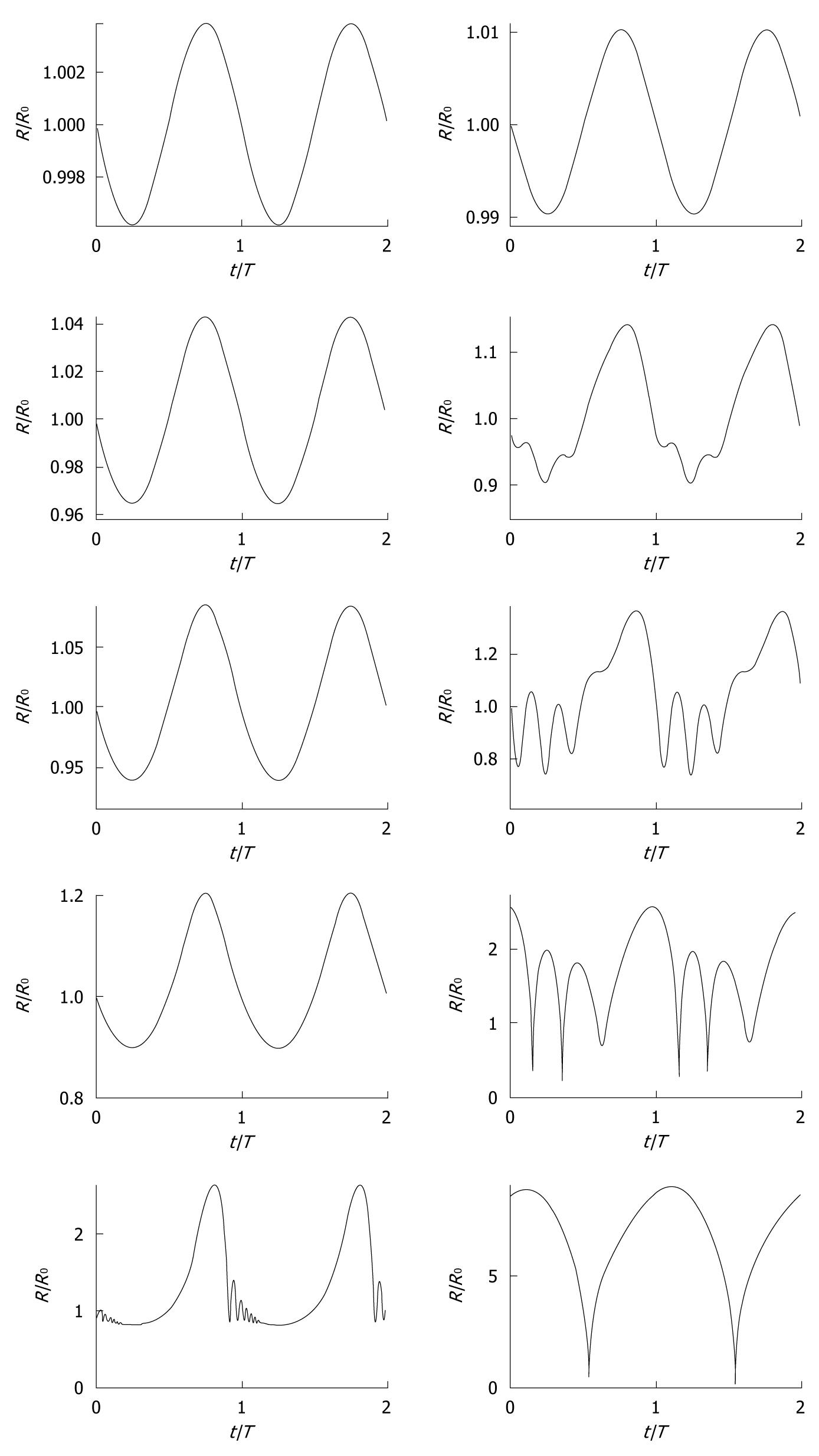
Figure 3 Simulated radius-time curves (radius R normalized with equilibrium radius R0, time t normalized with period T0) of ultrasound contrast microbubbles with 0.
55 μm (left column) and 2.3 μm (right column) equilibrium radii, respectively, modeled with a conservative Rayleigh-Plesset equation[3], using a conservative shell stiffness parameter[148]. The modeled ultrasound field was a continuous sine wave with a frequency of 0.5 MHz and acoustic amplitudes corresponding to (top-bottom) mechanical index = 0.01, 0.10, 0.18, 0.35, and 0.80, similar to the experiments by Karshafian et al[92].
- Citation: Postema M, Gilja OH. Contrast-enhanced and targeted ultrasound. World J Gastroenterol 2011; 17(1): 28-41
- URL: https://www.wjgnet.com/1007-9327/full/v17/i1/28.htm
- DOI: https://dx.doi.org/10.3748/wjg.v17.i1.28


