Copyright
©2010 Baishideng.
World J Gastroenterol. Feb 7, 2010; 16(5): 538-546
Published online Feb 7, 2010. doi: 10.3748/wjg.v16.i5.538
Published online Feb 7, 2010. doi: 10.3748/wjg.v16.i5.538
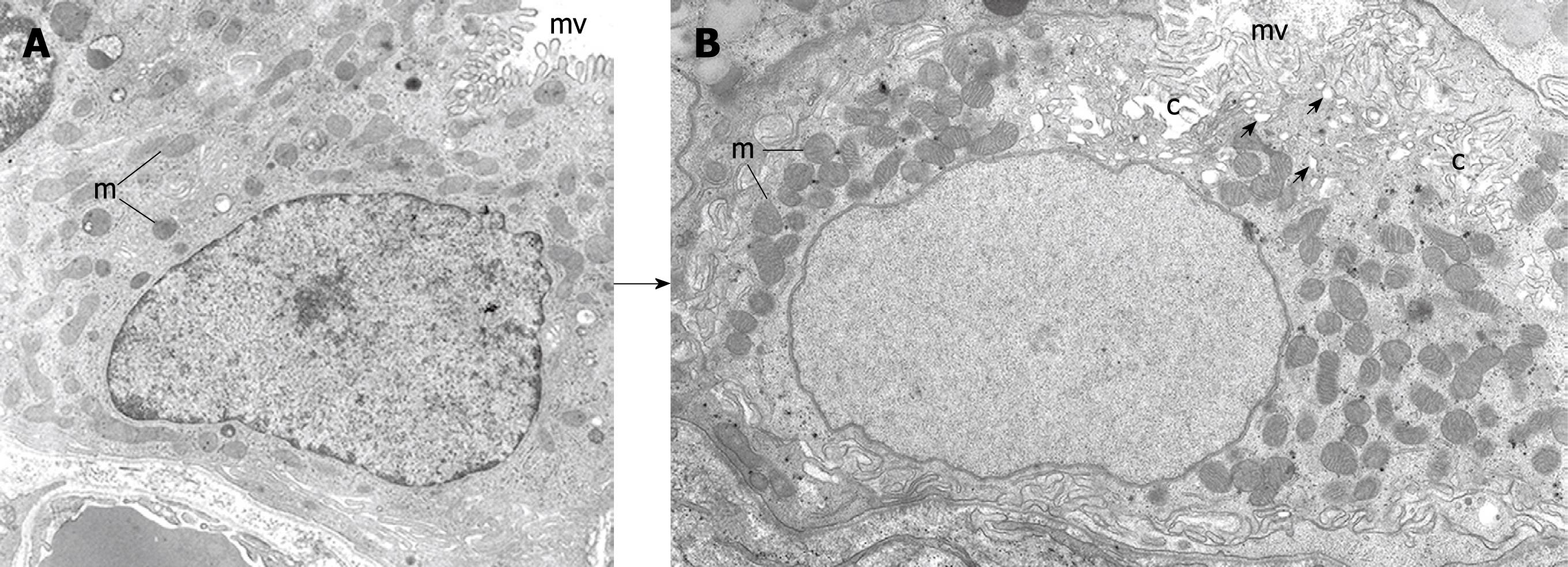
Figure 3 Electron micrographs depicting 2 stages of the differentiating preparietal cells as seen in the human stomach.
Original magnification, × 12 000. A: Preparietal cell showing relatively long numerous microvilli (mv). The mitochondria (m) are relatively few and small; B: Preparietal cell showing an incipient canaliculus (c) and relatively numerous mitochondria (m) which appear larger than those in A. The apical cytoplasm shows few tubulovesicles (small arrows) and long numerous microvilli (mv). A is reproduced from Karam et al[4] with permission from Wiley-Blackwell/AlphaMed press.
- Citation: Karam SM. A focus on parietal cells as a renewing cell population. World J Gastroenterol 2010; 16(5): 538-546
- URL: https://www.wjgnet.com/1007-9327/full/v16/i5/538.htm
- DOI: https://dx.doi.org/10.3748/wjg.v16.i5.538


