Copyright
©2010 Baishideng.
World J Gastroenterol. Aug 7, 2010; 16(29): 3664-3673
Published online Aug 7, 2010. doi: 10.3748/wjg.v16.i29.3664
Published online Aug 7, 2010. doi: 10.3748/wjg.v16.i29.3664
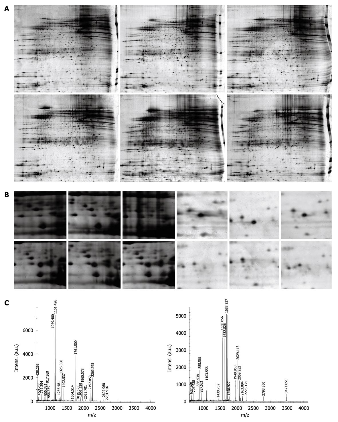
Figure 1 Detection and analysis of differentially expressed proteins in varioliform gastritis.
A: Representative 2-DE images of matched varioliform gastritis and normal gastric mucosa tissue. The proteins expressed in varioliform gastritis were compared with those expressed in matched normal tissue. The protein spots that showed more than 5-fold differential expression in at least nine cases were taken as differentially expressed candidates. Of 21 differentially expressed protein spots, 18 were identified by mass spectrometry (MS) (protein nomenclature can be seen in Table 2); B: The magnified regions of the 2-DE gel of upregulated thioredoxin domain-containing protein 5 (TXNDC5) (left) and downregulated phosphatidylethanolamine-binding protein 1 (PEBP1) (right) in varioliform gastritis, compared with normal tissue; C: MS of in-gel trypsin digests of these proteins and analysis of the depicted peptide spectrum resulted in the identification of TXNDC5 (left) and PEBP1 (right).
-
Citation: Zhang L, Hou YH, Wu K, Zhai JS, Lin N. Proteomic analysis reveals molecular biological details in varioliform gastritis without
Helicobacter pylori infection. World J Gastroenterol 2010; 16(29): 3664-3673 - URL: https://www.wjgnet.com/1007-9327/full/v16/i29/3664.htm
- DOI: https://dx.doi.org/10.3748/wjg.v16.i29.3664


