Copyright
©2010 Baishideng.
World J Gastroenterol. May 28, 2010; 16(20): 2476-2483
Published online May 28, 2010. doi: 10.3748/wjg.v16.i20.2476
Published online May 28, 2010. doi: 10.3748/wjg.v16.i20.2476
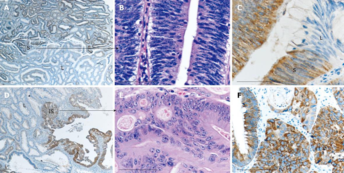
Figure 2 Heterogeneity of AMACR staining may be observed in different dysplastic areas within the same lesion.
Case 47, as listed in Figure 1A, is shown. This case contained areas of mild/moderate [low-grade (L)] and severe (S) dysplasia, as well as carcinoma in situ (IS) elements. Severely dysplastic (A-C) and carcinoma in situ areas (D-F) are strongly positive for AMACR, while mild/moderate dysplastic areas are negative. Histological distinction between severe dysplasia and carcinoma in situ is apparent in B and E, respectively (HE staining). Bars: 100 microns.
- Citation: Lakis S, Papamitsou T, Panagiotopoulou C, Kotakidou R, Kotoula V. AMACR is associated with advanced pathologic risk factors in sporadic colorectal adenomas. World J Gastroenterol 2010; 16(20): 2476-2483
- URL: https://www.wjgnet.com/1007-9327/full/v16/i20/2476.htm
- DOI: https://dx.doi.org/10.3748/wjg.v16.i20.2476


