Copyright
©2009 The WJG Press and Baishideng.
World J Gastroenterol. Mar 7, 2009; 15(9): 1057-1064
Published online Mar 7, 2009. doi: 10.3748/wjg.15.1057
Published online Mar 7, 2009. doi: 10.3748/wjg.15.1057
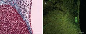
Figure 5 Histology and cytokeratin-19 immune staining of the liver at the boundary between the growing edge of the liver and the inactivated omentum at day 3 or 7 after injury.
A: Trichrome stained section showing the adherent omental tissue with a thinner interlying tissue (blue stained) than that seen in the activated omentum group (Figure 3 for comparison); B: Cytokeratin-19 positive bile ducts were seen in the interlying tissue (same section as A) although these were much less frequent than those seen in the activated omentum group (Figure 4 for comparison). The horizontal white bar in pictures represents 100 &mgr;m.
- Citation: Singh AK, Pancholi N, Patel J, Litbarg NO, Gudehithlu KP, Sethupathi P, Kraus M, Dunea G, Arruda JA. Omentum facilitates liver regeneration. World J Gastroenterol 2009; 15(9): 1057-1064
- URL: https://www.wjgnet.com/1007-9327/full/v15/i9/1057.htm
- DOI: https://dx.doi.org/10.3748/wjg.15.1057


