Copyright
©2009 The WJG Press and Baishideng.
World J Gastroenterol. Feb 7, 2009; 15(5): 570-577
Published online Feb 7, 2009. doi: 10.3748/wjg.15.570
Published online Feb 7, 2009. doi: 10.3748/wjg.15.570
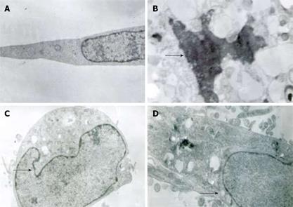
Figure 2 Mesothelial cells under electron microscope after incubation with and without SF-CM.
A: Normal nuclei and endoplasmic reticulum of control cells; B: Condensation of nuclear chromatin (arrow in Figure 2B); C: Wrinkling of nuclear membrane of cells after treatment with Astragalus injection (arrow in Figure 2C); D: Dilated endoplasmic reticulum (arrow in Figure 2D).
- Citation: Na D, Liu FN, Miao ZF, Du ZM, Xu HM. Astragalus extract inhibits destruction of gastric cancer cells to mesothelial cells by anti-apoptosis. World J Gastroenterol 2009; 15(5): 570-577
- URL: https://www.wjgnet.com/1007-9327/full/v15/i5/570.htm
- DOI: https://dx.doi.org/10.3748/wjg.15.570


