Copyright
©2009 The WJG Press and Baishideng.
World J Gastroenterol. Dec 14, 2009; 15(46): 5776-5783
Published online Dec 14, 2009. doi: 10.3748/wjg.15.5776
Published online Dec 14, 2009. doi: 10.3748/wjg.15.5776
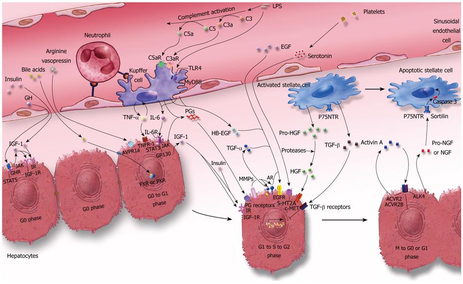
Figure 1 Scheme depicting the network of signaling among hepatic cells and blood during liver regeneration.
After liver injury such as PH, gut-derived factors such as LPS reach the liver through the portal blood supply. LPS activate the complement system, which releases the anaphylatoxins C3a and C5a. LPS, C3a and C5a activate Kupffer cells through TLR4, C3aR and C5aR, and further lead to production of TNF-α and IL-6. These factors are involved in priming the hepatocytes from G0 to G1 phase. Insulin, GH, bile acids, AVP, platelet-derived serotonin, and EGF from the blood, cooperating with PGs, HB-EGF, HGF, as well as IGF-1, TGF-α and AR from different hepatic cells, promote hepatocyte transition from G1, through S and G2, to the M phase of the cell cycle. TGF-β produced mainly by HSCs inhibits G1 to S phase transition of hepatocytes, and TGF-β signalling is blocked during the proliferative phase. Pro-NGF or NGF produced by the neighboring regenerating hepatocytes promotes the termination of the activated state of HSCs by pro-NGF- or NGF-induced apoptotic pathways. When the liver mass is restored to its normal volume, the increased signaling by activin A, apoptosis and other factors may promote termination of liver regeneration. The prototype of this figure is originated from reference[43].
- Citation: Zheng ZY, Weng SY, Yu Y. Signal molecule-mediated hepatic cell communication during liver regeneration. World J Gastroenterol 2009; 15(46): 5776-5783
- URL: https://www.wjgnet.com/1007-9327/full/v15/i46/5776.htm
- DOI: https://dx.doi.org/10.3748/wjg.15.5776


