Copyright
©2009 The WJG Press and Baishideng.
World J Gastroenterol. Nov 7, 2009; 15(41): 5165-5175
Published online Nov 7, 2009. doi: 10.3748/wjg.15.5165
Published online Nov 7, 2009. doi: 10.3748/wjg.15.5165
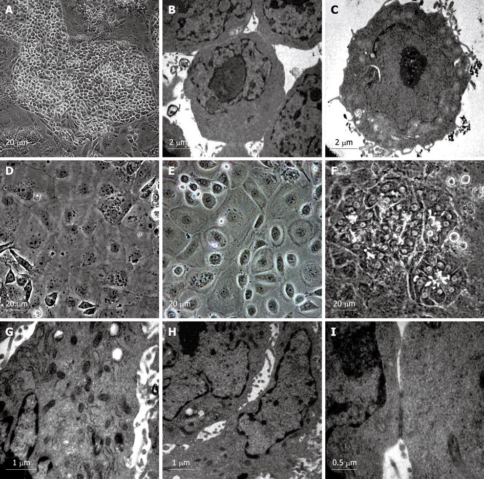
Figure 2 Morphological observation of the differentiated cells.
A: Morphology of hepatic progenitor cells induced by VPA; B: Electron microscopic observation of the undifferentiated ES cells (control); C: Electron microscopic observation of the hepatic progenitor cells differentiated from ES cells; D, E: Morphological observation of typical hepatocytes differentiated from hepatic progenitor cells with flattened and cuboidal morphology, and acquired abundant granules in the cytoplasm; F: Morphological observation of another kind of hepatocyte-like cells differentiated from hepatic progenitor cells with rising and piled morphology, dark cytoplasm and light nuclei, and bile canaliculi-like structures found between these cells; G: Electron microscopic observation of the differentiated hepatocytes with abundant mitochondria in their cytoplasm; H: Electron microscopic observation of the bile canaliculi between adjacent cells as mentioned above (Figure 1F); I: The bile canaliculi between adjacent cells sealed with tight junctions.
- Citation: Dong XJ, Zhang GR, Zhou QJ, Pan RL, Chen Y, Xiang LX, Shao JZ. Direct hepatic differentiation of mouse embryonic stem cells induced by valproic acid and cytokines. World J Gastroenterol 2009; 15(41): 5165-5175
- URL: https://www.wjgnet.com/1007-9327/full/v15/i41/5165.htm
- DOI: https://dx.doi.org/10.3748/wjg.15.5165


