Copyright
©2009 The WJG Press and Baishideng.
World J Gastroenterol. Aug 7, 2009; 15(29): 3611-3620
Published online Aug 7, 2009. doi: 10.3748/wjg.15.3611
Published online Aug 7, 2009. doi: 10.3748/wjg.15.3611
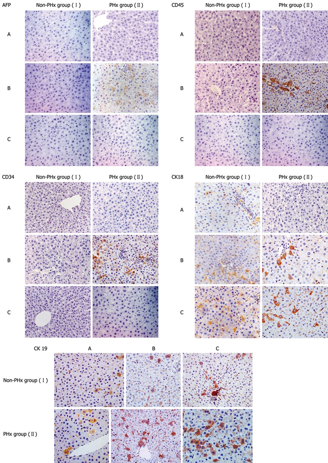
Figure 3 Representative immunohistochemistric analysis of phenotypic changes of donor-derived human cells in HRC liver during PHx-induced liver regeneration.
Seventy percent partial hepatectomy (PHx), a procedure that removes 70% of the liver, is regarded as the preferred in vivo method to study liver growth due to its synchronized growth response. In the representative chimeric livers of animals 5 and 6 (Table 1), donor-derived human cells with different cellular phenotypes were detected by IHC for different human markers (AFP, CD34, CD45, CK18 and CK19). Minimal liver tissues used for IHC were collected before PHx, and after 10 and 20 d of PHx. Additionally, the untransplanted control group was also set up when we performed this experiment (data not shown). The results demonstrate that CD45+, CK18+, AFP+, CD34+ and CK19+ cells could not be detected in liver sections from untransplanted control rats by human-specific IHC, suggesting that human-specific IHC can show human specificity of staining (data not shown). PHx: 70% partial hepatectomy, I: Non-PHx group; II: PHx group. A: before PHx; B: 10 d after PHx; C: 20 d after PHx.
- Citation: Sun Y, Xiao D, Li HA, Jiang JF, Li Q, Zhang RS, Chen XG. Phenotypic changes of human cells in human-rat liver during partial hepatectomy-induced regeneration. World J Gastroenterol 2009; 15(29): 3611-3620
- URL: https://www.wjgnet.com/1007-9327/full/v15/i29/3611.htm
- DOI: https://dx.doi.org/10.3748/wjg.15.3611


