Copyright
©2009 The WJG Press and Baishideng.
World J Gastroenterol. Jun 28, 2009; 15(24): 3038-3045
Published online Jun 28, 2009. doi: 10.3748/wjg.15.3038
Published online Jun 28, 2009. doi: 10.3748/wjg.15.3038
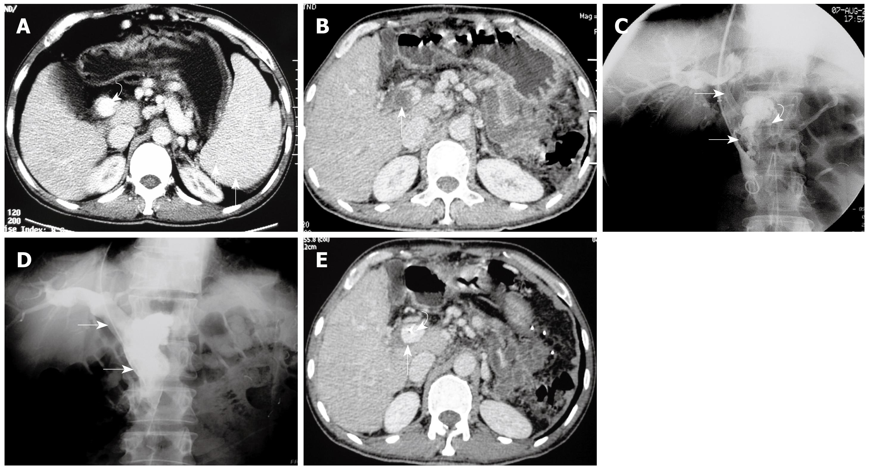
Figure 1 Case 1: A 40-year-old man with low-grade fever, abdominal pain, distension, and nausea for 12 d.
He had undergone splenectomy 4 wk previously. A: Selected axial image from before open splenectomy contrast-enhanced CT shows splenomegaly (arrows) and patent portal vein (curved arrow); B: Selected axial image from admission contrast-enhanced CT, on postoperative day 28, shows massive thrombus within the PV (arrow); C: Pre-treatment direct venography via transjugular approach access to the portal vein shows massive thrombosis of the PV extending into the SMV (arrows). Note the stump of the splenic vein (curved arrow); D: Follow-up direct portal venography via the infusion catheter, obtained 5 d after the catheter infusion of thrombolytics, shows patency of the main PV-SMV (arrows); E: CT image at the same level as in Figure 1B obtained 5 d after the interventional procedure, before the infusion catheter removal, shows the wide patent main PV (arrow). Note the infusion catheter within the PV (curved arrow).
- Citation: Wang MQ, Lin HY, Guo LP, Liu FY, Duan F, Wang ZJ. Acute extensive portal and mesenteric venous thrombosis after splenectomy: Treated by interventional thrombolysis with transjugular approach. World J Gastroenterol 2009; 15(24): 3038-3045
- URL: https://www.wjgnet.com/1007-9327/full/v15/i24/3038.htm
- DOI: https://dx.doi.org/10.3748/wjg.15.3038


