Copyright
©2009 The WJG Press and Baishideng.
World J Gastroenterol. Jan 14, 2009; 15(2): 160-168
Published online Jan 14, 2009. doi: 10.3748/wjg.15.160
Published online Jan 14, 2009. doi: 10.3748/wjg.15.160
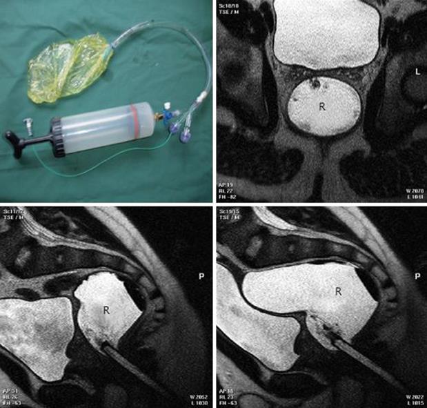
Figure 8 Stepwise distension of water filled balloon with simultaneous MRI and pressure recording.
The rectal probe (upper left) allows rectal water distension and pressure measurement. MRI shows the distended water-filled bag in the rectum (R). The sagittal MRI (lower panel) shows the distension, elongation and relation to neighbouring structures at 100 mL and 300 mL inside the bag. Modified from [50].
- Citation: Frøkjær JB, Drewes AM, Gregersen H. Imaging of the gastrointestinal tract-novel technologies. World J Gastroenterol 2009; 15(2): 160-168
- URL: https://www.wjgnet.com/1007-9327/full/v15/i2/160.htm
- DOI: https://dx.doi.org/10.3748/wjg.15.160


