Copyright
©2009 The WJG Press and Baishideng.
World J Gastroenterol. Jan 14, 2009; 15(2): 160-168
Published online Jan 14, 2009. doi: 10.3748/wjg.15.160
Published online Jan 14, 2009. doi: 10.3748/wjg.15.160
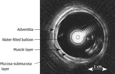
Figure 2 Cross-sectional endosonographic image of the distended distal oesophagus allows identification of three oesophageal sub-layers, i.
e. mucosa-submucosa, muscle and adventitia. The white shadows inside the water-filled bag (4-5 o’clock) represent artefacts due to convulsions of the water-filled balloon, which results in reduced image quality at low degrees of distension. Modified from [17].
- Citation: Frøkjær JB, Drewes AM, Gregersen H. Imaging of the gastrointestinal tract-novel technologies. World J Gastroenterol 2009; 15(2): 160-168
- URL: https://www.wjgnet.com/1007-9327/full/v15/i2/160.htm
- DOI: https://dx.doi.org/10.3748/wjg.15.160


