Copyright
©2007 Baishideng Publishing Group Co.
World J Gastroenterol. Sep 21, 2007; 13(35): 4673-4689
Published online Sep 21, 2007. doi: 10.3748/wjg.v13.i35.4673
Published online Sep 21, 2007. doi: 10.3748/wjg.v13.i35.4673
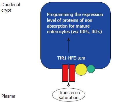
Figure 1 The duodenal “crypt cell hypothesis” of HFE function[36].
HFE at the basolateral membrane of duodenal crypt cells co-localizes with β2-microglobulin and TfR1. The saturation of circulating transferrin, which reflects body iron stores, was proposed to be “sensed” by the TfR1-HFE-β2-microglobulin complex, through transferrin-mediated endocytosis. Wild type HFE was proposed to facilitate transferrin-mediated iron uptake. In haemochromatosis, deficiency of functional HFE would therefore decrease the iron pool within the crypt cell, despite increased body iron stores. This would increase the activity of iron responsive proteins (IRPs), leading to increased expression of genes involved in iron absorption, such as DMT1 and ferroportin. This could contribute to the iron overload seen in HH[27,34,35]. However, recent evidence suggests that HFE may play more important roles in influencing iron metabolism in the liver.
-
Citation: Sebastiani G, Walker AP.
HFE gene in primary and secondary hepatic iron overload. World J Gastroenterol 2007; 13(35): 4673-4689 - URL: https://www.wjgnet.com/1007-9327/full/v13/i35/4673.htm
- DOI: https://dx.doi.org/10.3748/wjg.v13.i35.4673


