Copyright
©2007 Baishideng Publishing Group Co.
World J Gastroenterol. Jun 21, 2007; 13(23): 3255-3258
Published online Jun 21, 2007. doi: 10.3748/wjg.v13.i23.3255
Published online Jun 21, 2007. doi: 10.3748/wjg.v13.i23.3255
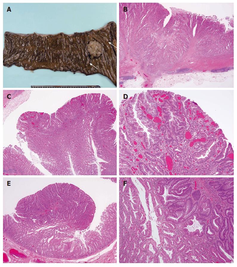
Figure 4 A: Resected ascending colon.
A cancer, 2.8 cm × 3.8 cm in size, and multiple small polyps are seen. The two largest polyps are 13 mm × 8 mm and 11 mm × 4 mm in size, respectively (arrows); B: Well to moderately differentiated adenocarcinoma (HE, × 20). Atypical duct formation and cribriform pattern can be seen. The tumor infiltrates the muscular layer and reaches the subserosal layer without serosal exposure; C: A small area from the periphery of the adenocarcinoma (HE, × 20). The lower part is the hyperplastic polyp component, and the upper part is the adenocarcinoma component; D: The left side of Figure 4C (HE, × 100). A small adenoma-like component is shown in the border between the hyperplastic component and the adenocarcinoma component; E: The combined adenoma-hyperplastic polyp (HE, × 20). The polyp is 1 cm in size. The upper part is the adenoma component, which primarily consists of a tubular adenoma with mild cellular atypia, the lower part is the hyperplastic polyp component; F: A higher magnification of Figure 4E (HE, × 100). A small amount of serrated structure with mild atypia is seen at the border of the two components.
- Citation: Kurobe M, Abe K, Kinoshita N, Anami M, Tokai H, Ryu Y, Wen CY, Kanematsu T, Hayashi T. Hyperplastic polyposis associated with two asynchronous colon cancers. World J Gastroenterol 2007; 13(23): 3255-3258
- URL: https://www.wjgnet.com/1007-9327/full/v13/i23/3255.htm
- DOI: https://dx.doi.org/10.3748/wjg.v13.i23.3255


