Copyright
©2007 Baishideng Publishing Group Co.
World J Gastroenterol. Apr 21, 2007; 13(15): 2160-2165
Published online Apr 21, 2007. doi: 10.3748/wjg.v13.i15.2160
Published online Apr 21, 2007. doi: 10.3748/wjg.v13.i15.2160
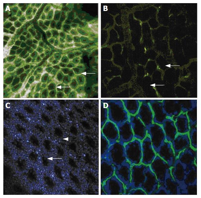
Figure 3 A: Intravenous fluorescein sodium gives a thorough impression of the colonic mucosa.
The crypt lumina are still visible within, although at this tissue depth (about 150 μm), sub cellular details such as mucin in goblet cells can no longer be displayed. Larger vessels drain and feed the capillaries, which are rendered as white bands (arrows) surrounding the tightly packed crypts; B: After intravenous injection of FITC-labelled L. esculentum lectin, fluorescent labelling of the endothelial cells results in a selective staining of the capillary walls of the colonic mucosa. The enterocytes and goblet cells of the crypt itself (arrows) are not visualized; C: After injection of acriflavine, visualization of the epithelial cell nuclei renders the regular pattern of crypts in normal colonic mucosa. Cells are captured either en face (in-between crypts, arrow) or transverse (in the pits, arrowhead); D: For the bench-top confocal correlate, an ex vivo nuclear counter stain was performed with Hoechst blue, rendering the circular arrangement of basally oriented nuclei of the colonic crypts. The labelling of vessel walls was performed in vivo. This double staining corresponds to an ex vivo overlay of Figure 3B and C.
-
Citation: Goetz M, Memadathil B, Biesterfeld S, Schneider C, Gregor S, Galle PR, Neurath MF, Kiesslich R.
In vivo subsurface morphological and functional cellular and subcellular imaging of the gastrointestinal tract with confocal mini-microscopy. World J Gastroenterol 2007; 13(15): 2160-2165 - URL: https://www.wjgnet.com/1007-9327/full/v13/i15/2160.htm
- DOI: https://dx.doi.org/10.3748/wjg.v13.i15.2160


