Copyright
©2007 Baishideng Publishing Group Co.
World J Gastroenterol. Apr 21, 2007; 13(15): 2160-2165
Published online Apr 21, 2007. doi: 10.3748/wjg.v13.i15.2160
Published online Apr 21, 2007. doi: 10.3748/wjg.v13.i15.2160
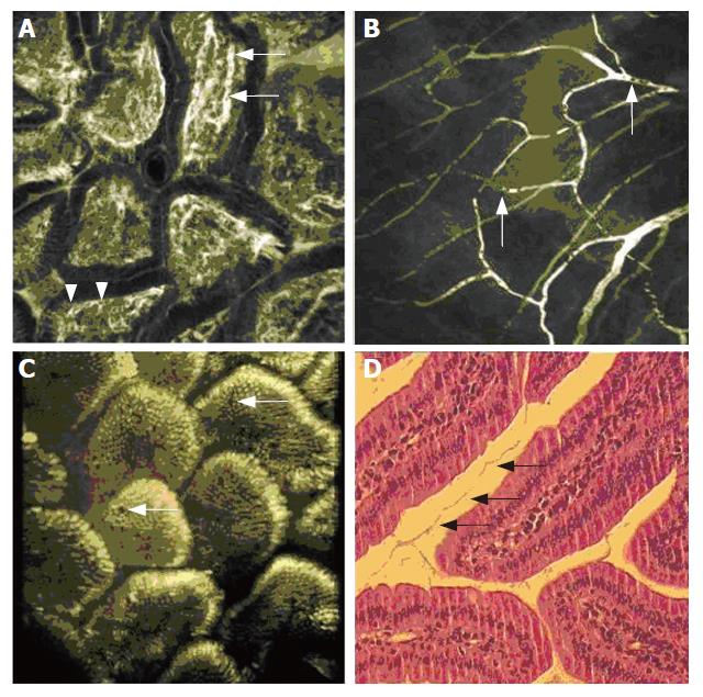
Figure 2 A: Duodenal villi after intravenous injection of FITC-labelled dextran.
Capillaries are brightly contrasted, while blood cells are rendered as black dots within the lumen (arrow). Vessel loops are situated close to the clear cut basal border of columnar epithelial cells (arrow head); B: After acriflavine application, the regular distribution of enterocytes on the villi is visualized. Interspersed are multiple goblet cells in which the mucin is only weakly stained (arrows). A still from a three-dimensional reconstruction of 21 serial transverse sections is rendered, the full reconstruction is given as a video file as Suppl. Figure 1; C: In the corresponding H&E stained specimen, the villi seem to be set further apart. The mucus filaments (arrows) probably indicate the prior extent of the villi, illustrating the range of shrinking artifacts upon fixation; D: In the fine vessel structure of the small intestinal meso, blood plasma is brightly contrasted after injection of FITC-labeled dextran. Black inclusions within the plasma flow correspond to unstained blood cells (arrows).
-
Citation: Goetz M, Memadathil B, Biesterfeld S, Schneider C, Gregor S, Galle PR, Neurath MF, Kiesslich R.
In vivo subsurface morphological and functional cellular and subcellular imaging of the gastrointestinal tract with confocal mini-microscopy. World J Gastroenterol 2007; 13(15): 2160-2165 - URL: https://www.wjgnet.com/1007-9327/full/v13/i15/2160.htm
- DOI: https://dx.doi.org/10.3748/wjg.v13.i15.2160


