Copyright
©2007 Baishideng Publishing Group Co.
World J Gastroenterol. Mar 14, 2007; 13(10): 1477-1486
Published online Mar 14, 2007. doi: 10.3748/wjg.v13.i10.1477
Published online Mar 14, 2007. doi: 10.3748/wjg.v13.i10.1477
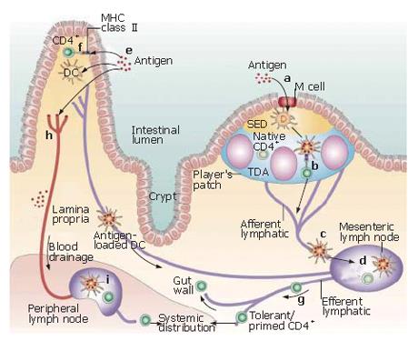
Figure 2 Antigen uptake and recognition by CD4+ T cells in the intestine.
Antigen may enter through the microfold (M) cells in the follicle-associated epithelium (FAE) (a), and after transfer to local dendritic cells (DCs), might then be presented directly to T cells in the Peyer’s patch (b). Alternatively, antigen or antigen-loaded DCs from the Peyer’s patch may gain access to draining lymph (c), with subsequent T-cell recognition in the mesenteric lymph nodes (MLNs) (d). A similar process of antigen or antigen-presenting cell (APC) dissemination to MLNs may occur if antigen enters through the epithelium covering the villus lamina propria (e), but in this case, there is the further possibility that MHC class II+ enterocytes may act as local APCs (f). In all cases, the antigen-responsive CD4+ T cells acquire expression of the α4β7 integrin and the chemokine receptor CCR9, leave the MLN in the efferent lymph (g) and after entering the bloodstream through the thoracic duct, exit into the mucosa through vessels in the lamina propria. T cells which have recognized antigen first in the MLN may also disseminate from the bloodstream throughout the peripheral immune system. Antigen may also gain direct access to the bloodstream from the gut (h) and interact with T cells in peripheral lymphoid tissues (i). (Adapted by permission from Macmillan Publishers Ltd: Nature Reviews Immunology[32], copyright 2003).
- Citation: Miller H, Zhang J, KuoLee R, Patel GB, Chen W. Intestinal M cells: The fallible sentinels? World J Gastroenterol 2007; 13(10): 1477-1486
- URL: https://www.wjgnet.com/1007-9327/full/v13/i10/1477.htm
- DOI: https://dx.doi.org/10.3748/wjg.v13.i10.1477


