Copyright
©2006 Baishideng Publishing Group Co.
World J Gastroenterol. Mar 7, 2006; 12(9): 1476-1478
Published online Mar 7, 2006. doi: 10.3748/wjg.v12.i9.1476
Published online Mar 7, 2006. doi: 10.3748/wjg.v12.i9.1476
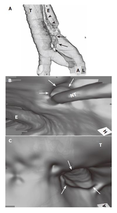
Figure 2 A: CT esophagography demonstrates communication between the middle intrathoracic esophagus and the distal trachea just proximal to the carina (arrows).
Note a distortion caused by tube inserted into the esophagus (arrowheads). T, trachea; E, esophagus. B: Virtual esophagoscopy shows the orifice of the fistula (arrows) and it is similar to that of (conventional) esophagoscopy (Figure 1). NT, naso-gastric tube; E, esophagus. C: Virtual bronchoscopy also demonstrates the orifice of the fistula (arrows). T, trachea.
- Citation: Nagata K, Kamio Y, Ichikawa T, Kadokura M, Kitami A, Endo S, Inoue H, Kudo SE. Congenital tracheoesophageal fistula successfully diagnosed by CT esophagography. World J Gastroenterol 2006; 12(9): 1476-1478
- URL: https://www.wjgnet.com/1007-9327/full/v12/i9/1476.htm
- DOI: https://dx.doi.org/10.3748/wjg.v12.i9.1476


