Copyright
©2006 Baishideng Publishing Group Co.
World J Gastroenterol. Sep 7, 2006; 12(33): 5320-5325
Published online Sep 7, 2006. doi: 10.3748/wjg.v12.i33.5320
Published online Sep 7, 2006. doi: 10.3748/wjg.v12.i33.5320
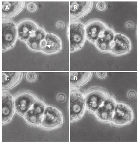
Figure 1 Representative phase contrast micrographs of an isolated hepatocyte triplet in the controls.
Figures A, B, C and D show a sequence of the bile canalicular contractions. A bile canaliculus indicated by an arrowhead started to contract (A) and completed contraction (D). Another canaliculus indicated by an arrow started its contraction (B) and completed contraction (C). These canalicular contractions were forceful and repetitive.
- Citation: Watanabe N, Kagawa T, Kojima SI, Takashimizu S, Nagata N, Nishizaki Y, Mine T. Taurolithocholate impairs bile canalicular motility and canalicular bile secretion in isolated rat hepatocyte couplets. World J Gastroenterol 2006; 12(33): 5320-5325
- URL: https://www.wjgnet.com/1007-9327/full/v12/i33/5320.htm
- DOI: https://dx.doi.org/10.3748/wjg.v12.i33.5320


