Copyright
©2006 Baishideng Publishing Group Co.
World J Gastroenterol. Jun 14, 2006; 12(22): 3512-3522
Published online Jun 14, 2006. doi: 10.3748/wjg.v12.i22.3512
Published online Jun 14, 2006. doi: 10.3748/wjg.v12.i22.3512
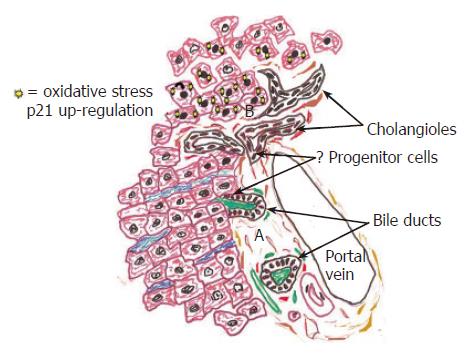
Figure 3 This diagram illustrates the peripheral aspects of the biliary tree, including the portal tract and periportal hepatic parenchyma in normal (A: bottom of figure) and in diseased livers (B: top of figure).
A: Rare liver progenitor, or stem cells, are thought to reside within the Canals of Herring, or the portion of the bile ductules that connects BECs to hepatocytes. The same close relationship between BEC, the arterial supply, and periductal myofibroblasts seen in the large ducts continues to the biliary tree periphery, as shown here. B: During chronic necro-inflammatory liver diseases a variety of insults, such as cholestasis[1], HCV replication[114,115], steatosis[2,116] copper deposition[117], and alcohol[118] can cause oxidative stress, preferentially in hepatocytes. The stress upregulates hepatocyte nuclear p21 expression (illustrated by solid nuclei at top of diagram), which in turn, inhibits hepatocytes proliferation and accentuates ductular reactions[1,2]. A close relationship between BEC and periductal myofibroblasts in the smallest bile ductules usually results in co-activation of these populations recognized as ductular reactions, which often precede the development of cirrhosis.
- Citation: Demetris AJ, III JGL, Specht S, Nozaki I. Biliary wound healing, ductular reactions, and IL-6/gp130 signaling in the development of liver disease. World J Gastroenterol 2006; 12(22): 3512-3522
- URL: https://www.wjgnet.com/1007-9327/full/v12/i22/3512.htm
- DOI: https://dx.doi.org/10.3748/wjg.v12.i22.3512


