Copyright
©2006 Baishideng Publishing Group Co.
World J Gastroenterol. May 28, 2006; 12(20): 3186-3195
Published online May 28, 2006. doi: 10.3748/wjg.v12.i20.3186
Published online May 28, 2006. doi: 10.3748/wjg.v12.i20.3186
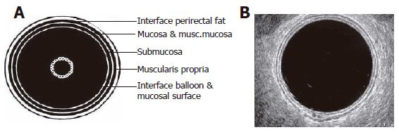
Figure 1 Rectal wall anatomy.
A: Schematic diagram of ERUS image; B: Actual image of normal ERUS[3].
- Citation: Balch GC, Meo AD, Guillem JG. Modern management of rectal cancer: A 2006 update. World J Gastroenterol 2006; 12(20): 3186-3195
- URL: https://www.wjgnet.com/1007-9327/full/v12/i20/3186.htm
- DOI: https://dx.doi.org/10.3748/wjg.v12.i20.3186


