Copyright
©2006 Baishideng Publishing Group Co.
World J Gastroenterol. May 21, 2006; 12(19): 2969-2978
Published online May 21, 2006. doi: 10.3748/wjg.v12.i19.2969
Published online May 21, 2006. doi: 10.3748/wjg.v12.i19.2969
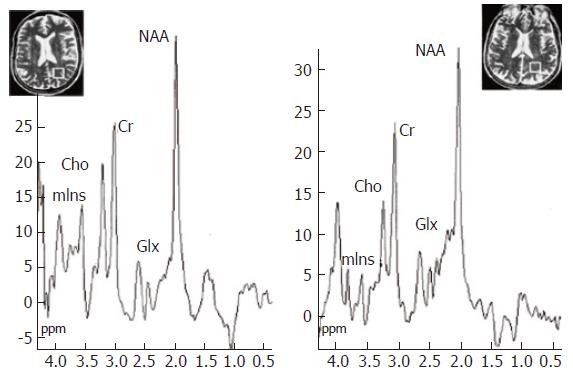
Figure 5 1H MR spectroscopy water-suppressed proton spectra of an 8 mL voxel located in the parietal region including predominantly normal appearing white matter (insert images show position of the voxel) in a healthy control subject (left) and in a cirrhotic patient (right), acquired using a STEAM pulse sequence [1600/20/30/256 (TR/TE/TM/acquisitions)].
The main resonances correspond to N-acetylaspartate (NAA, 2.0 ppm), glutamate/glutamine (Glx, 2.1-2.5 ppm), creatine/phosphocreatine (Cr, 3.02 ppm), choline-containing compounds (Cho, 3.2 ppm), and myo-Inositol (mIns, 3.55 ppm). Comparison of the spectra shows a decrease in Cho and mIns resonances with an increase in the glutamate/glutamine region in the cirrhotic patient. Reproduced with the permission of Springer Science and Business Media, from Cordoba et al 2002[28].
- Citation: Grover VB, Dresner MA, Forton DM, Counsell S, Larkman DJ, Patel N, Thomas HC, Taylor-Robinson SD. Current and future applications of magnetic resonance imaging and spectroscopy of the brain in hepatic encephalopathy. World J Gastroenterol 2006; 12(19): 2969-2978
- URL: https://www.wjgnet.com/1007-9327/full/v12/i19/2969.htm
- DOI: https://dx.doi.org/10.3748/wjg.v12.i19.2969


