Copyright
©2006 Baishideng Publishing Group Co.
World J Gastroenterol. Apr 14, 2006; 12(14): 2209-2216
Published online Apr 14, 2006. doi: 10.3748/wjg.v12.i14.2209
Published online Apr 14, 2006. doi: 10.3748/wjg.v12.i14.2209
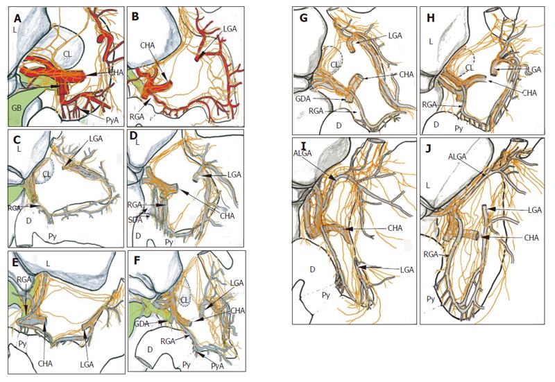
Figure 3 Diagram indicating the distribution in and around the cardia, the lesser curvature, the porta hepatica and the antro-pyloric region in 10 cadavers.
Among them, A and B are diagrams of Figures 1A and 2, respectively. Five specimens, B, D, F, I and J, showed hepatic divisions joining directly to the right gastric artery, while, for the other specimens, after joining to the proper hepatic or hepatic artery, the nerves sent off some offshoots to the right gastric artery, and innervated the pyloric region. ALGA, accessory left gastric artery; CHA, common hepatic artery; CL, caudal liver; GB, gallbladder; L, liver; LGA, left gastric artery; Py, pylorus; PyA, pyloric artery; RGA, right gastric artery; SDA, supra-duodenal artery.
- Citation: Yi SQ, Ru F, Ohta T, Terayama H, Naito M, Hayashi S, Buhe S, Yi N, Miyaki T, Tanaka S, Itoh M. Surgical anatomy of the innervation of pylorus in human and Suncus murinus, in relation to surgical technique for pylorus-preserving pancreaticoduodenectomy. World J Gastroenterol 2006; 12(14): 2209-2216
- URL: https://www.wjgnet.com/1007-9327/full/v12/i14/2209.htm
- DOI: https://dx.doi.org/10.3748/wjg.v12.i14.2209


