Copyright
©The Author(s) 2005.
World J Gastroenterol. Aug 7, 2005; 11(29): 4607-4609
Published online Aug 7, 2005. doi: 10.3748/wjg.v11.i29.4607
Published online Aug 7, 2005. doi: 10.3748/wjg.v11.i29.4607
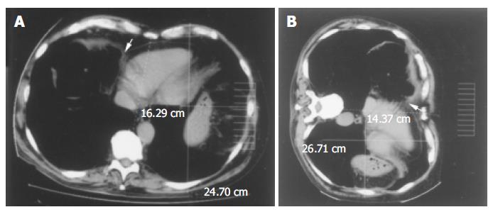
Figure 2 CT scan of the chest after air insufflation into the colon.
A: Supine position: the arrow shows the close vicinity of the hepatic flexure (asterisk) to the right side of the heart with the interposition of the diaphragm. However, the pericardium and the intestinal wall are clearly demarcated. The left hemithorax appears symmetrical compared to the right one; B: Left lateral decubitus: upon changing position the colon moves forward and medially, coming into contact with the vena cava and the right side of the heart (arrow). Compared to the supine position, the left hemithorax anteroposterior diameter is increased while the transverse diameter is decreased. This phenomenon may, at least in part, be associated to partial migration of the diaphragm and its attached organs and vessels toward the left side of the chest. This is also underlined by the change in the position of the heart in this radiogram in relation to the broken line (drawn from the sternum to the vertebra) compared to the radiogram in Figure 1. In this posture the patient complained of his typical precordial pain.
- Citation: Sorrentino D, Bazzocchi M, Badano L, Toso F, Giagu P. Heart-touching Chilaiditi’s syndrome. World J Gastroenterol 2005; 11(29): 4607-4609
- URL: https://www.wjgnet.com/1007-9327/full/v11/i29/4607.htm
- DOI: https://dx.doi.org/10.3748/wjg.v11.i29.4607


