Copyright
©The Author(s) 2005.
World J Gastroenterol. Aug 7, 2005; 11(29): 4490-4496
Published online Aug 7, 2005. doi: 10.3748/wjg.v11.i29.4490
Published online Aug 7, 2005. doi: 10.3748/wjg.v11.i29.4490
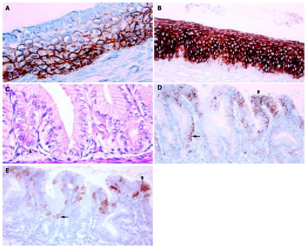
Figure 3 Tubular esophagus.
A: Esophageal ciliated epithelium at 20-wk GA. The immunostaining is for CK5 and is more intense in the basal and intermediate cell layers than in the superficial cell layer (OM ×400); B: Esophageal squamous epithelium in a 3-wk-old neonate. The immunostaining is for CK13 and is absent in the basal cell layer and diffuse moderate to strong in the intermediate and superficial cell layers (OM ×200); C: Esophageal simple columnar epithelium at 20-wk GA. To the left: parietal cells with roughly triangular shape and highly acidophilic cytoplasm (ESP, arrowhead). To the right: no discernible parietal cells are present (ESN, H&E, OM ×400); D: Esophageal simple columnar epithelium at 20-wk GA. The immunostaining is for CK7 and shows moderate positivity at the mucosal surface and deep in the glands (arrowhead and arrow, respectively, OM ×200); E: Esophageal simple columnar epithelium at 20-wk GA (same case as in Figure 3D). The immunostaining is for CK20 and shows patchy positivity of the superficial part of the mucosa (surface and pit epithelium: arrowhead and arrow, respectively, OM ×200).
- Citation: Hertogh GD, Eyken PV, Ectors N, Geboes K. On the origin of cardiac mucosa: A histological and immunohistoc-hemical study of cytokeratin expression patterns in the developing esophagogastric junction region and stomach. World J Gastroenterol 2005; 11(29): 4490-4496
- URL: https://www.wjgnet.com/1007-9327/full/v11/i29/4490.htm
- DOI: https://dx.doi.org/10.3748/wjg.v11.i29.4490


