Copyright
©The Author(s) 2005.
World J Gastroenterol. Jul 14, 2005; 11(26): 4061-4066
Published online Jul 14, 2005. doi: 10.3748/wjg.v11.i26.4061
Published online Jul 14, 2005. doi: 10.3748/wjg.v11.i26.4061
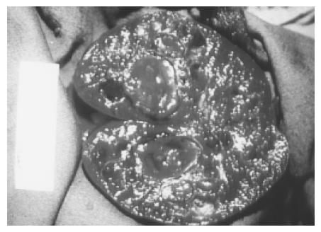
Figure 6 Large and multiple hemangiomas occupy the entire spleen.
The hypoechoic areas shown in Figure 5 are filled with blood clots and thrombosis. The 48-year-old male received splenectomy due to LUQ pain caused by venous thrombosis in spleen.
- Citation: Chen MJ, Huang MJ, Chang WH, Wang TE, Wang HY, Chu CH, Lin SC, Shih SC. Ultrasonography of splenic abnormalities. World J Gastroenterol 2005; 11(26): 4061-4066
- URL: https://www.wjgnet.com/1007-9327/full/v11/i26/4061.htm
- DOI: https://dx.doi.org/10.3748/wjg.v11.i26.4061


