Copyright
©2009 The WJG Press and Baishideng.
World J Gastroenterol. Oct 7, 2009; 15(37): 4617-4626
Published online Oct 7, 2009. doi: 10.3748/wjg.15.4617
Published online Oct 7, 2009. doi: 10.3748/wjg.15.4617
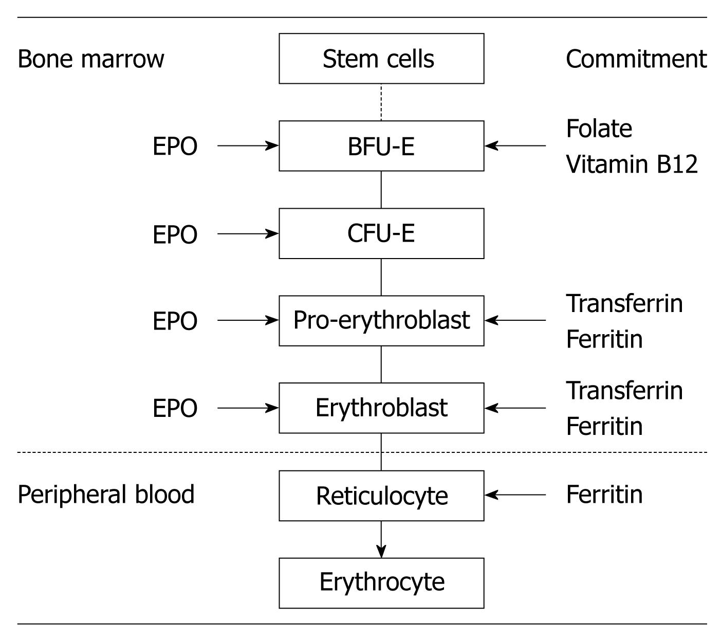
Figure 1 Major stages of human erythropoiesis showing the point of commitment, the period of EPO dependence and the requirements for essential nutrients.
BFU-E: Burst-forming unit-erythroid; CFU-E: Colony-forming unit-erythroid; EPO: Erythropoietin.
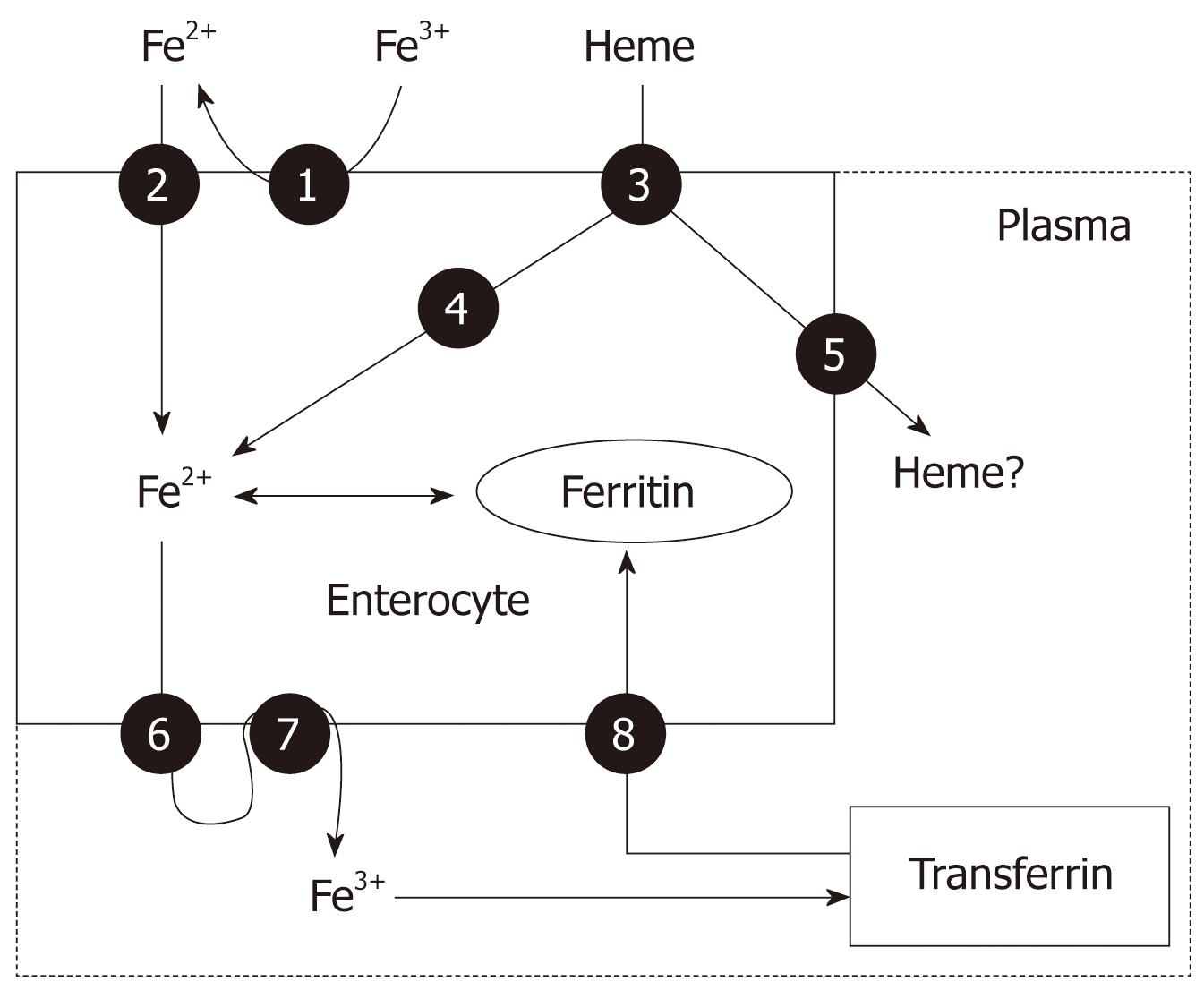
Figure 2 Main pathways of iron absorption by enterocytes in mammals.
1: Ferrireductase; 2: Divalent metal transporter 1 (DMT-1); 3: Heme protein carrier 1 (HPC1); 4: Heme oxygenase; 5: Heme exporter; 6: Ferroportin (Ireg-1); 7: Hephaestin; 8: Transferrin receptor-1 (TfR1) (for details see Table 1).
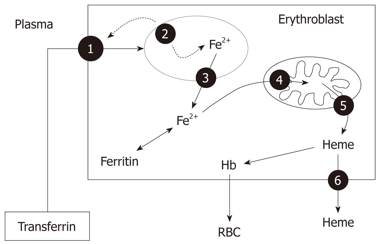
Figure 3 Main pathways of iron utilization by erythroblasts in mammals.
1: TfR1; 2: Diferric-transferrin-TfR1 complex; 3: Natural resistance macrophage protein (NRAMP-1); 4: Mitoferrin; 5: Mitochondrial heme exporter (Abcb6); 6: Heme exporter (FLVCR, Bcrp/Abcg2) (for details see Table 1).
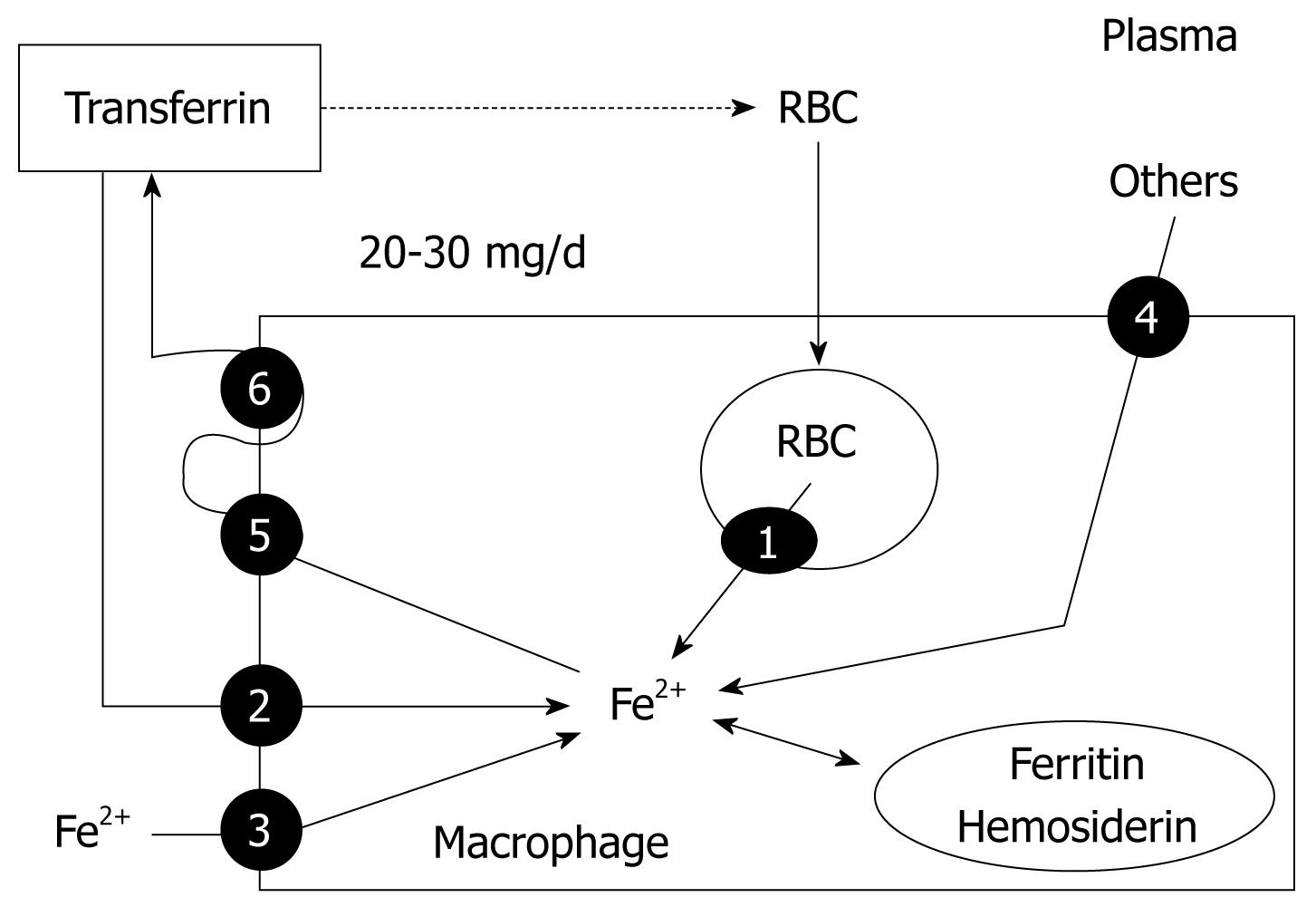
Figure 4 Main pathways of iron storage and exportation by macrophages in mammals.
1: NRAMP-1; 2: TfR1; 3: DMT-1; 4: Others: others: bacteria, lactoferrin, hemoglobin-haptoglobin, heme-hemopexin; 5: Ferroportin (Ireg-1); 6: Hephaestin (for details see Table 1).
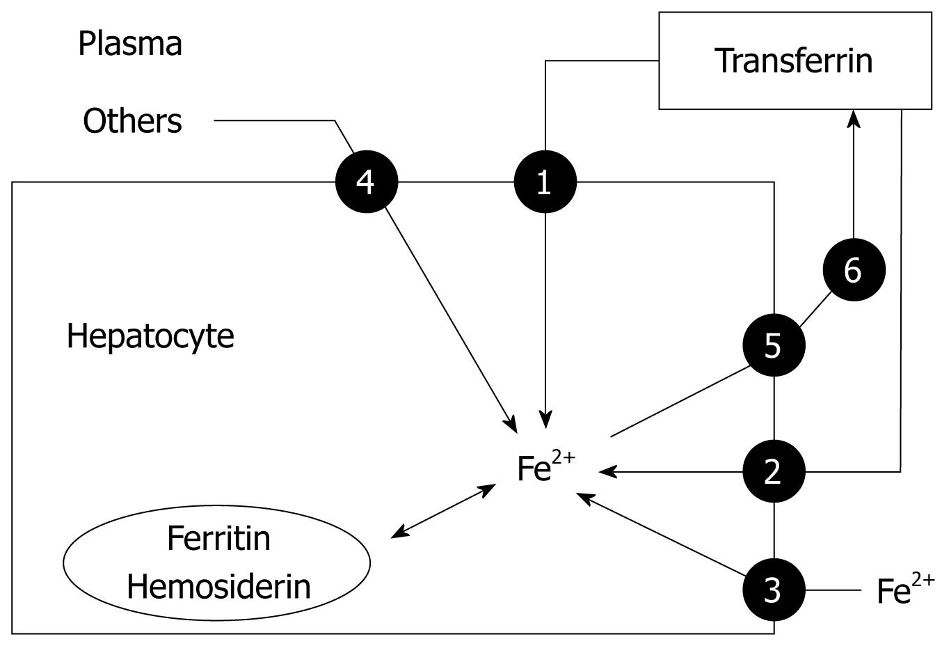
Figure 5 Main pathways of iron storage and exportation by hepatocytes in mammals.
1: TfR1; 2: TfR2; 3: DMT-1; 4: Others: hemoglobin, heme, ferritin; 5: Ferroportin (Ireg-1); 6: Ceruloplasmin (for details see Table 1).
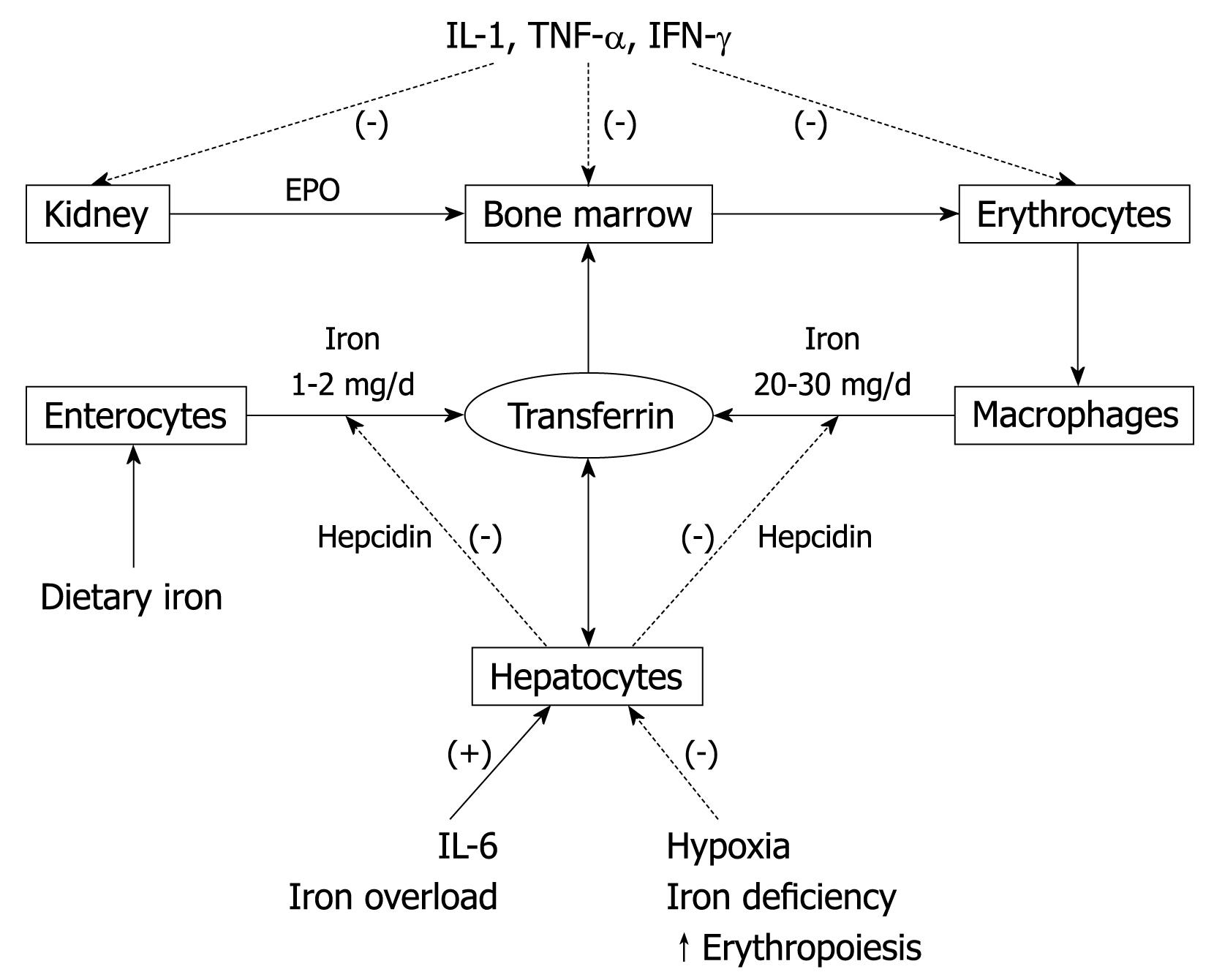
Figure 6 Effects of inflammation on erythropoiesis and iron homeostasis in mammals.
(-): Negative effect; (+): Positive effect.
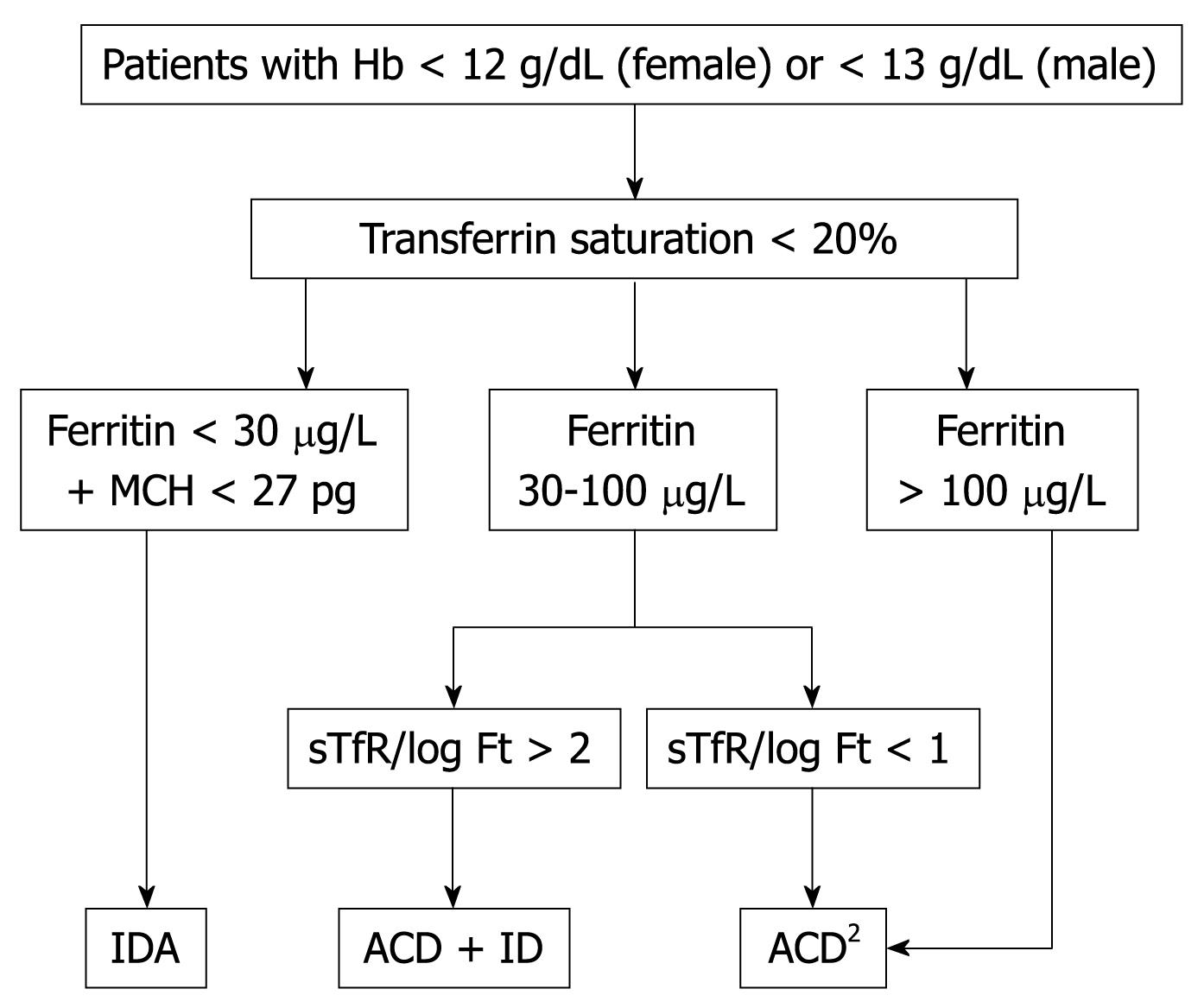
Figure 7 A simplified algorithm for the diagnosis of IDA (modified from Weiss et al[19]).
- Citation: Muñoz M, Villar I, García-Erce JA. An update on iron physiology. World J Gastroenterol 2009; 15(37): 4617-4626
- URL: https://www.wjgnet.com/1007-9327/full/v15/i37/4617.htm
- DOI: https://dx.doi.org/10.3748/wjg.15.4617


