Copyright
©2008 The WJG Press and Baishideng.
World J Gastroenterol. May 7, 2008; 14(17): 2731-2739
Published online May 7, 2008. doi: 10.3748/wjg.14.2731
Published online May 7, 2008. doi: 10.3748/wjg.14.2731
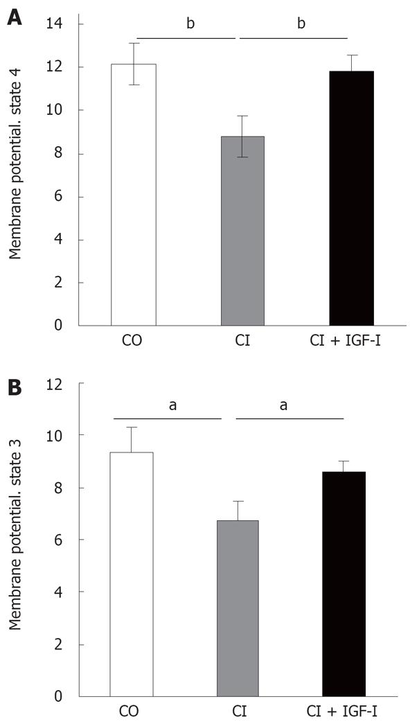
Figure 2 Mitochondrial Membrane Potential (MMP): the MMP was evaluated by flow cytometry with Rhodamine 123 under respiratory conditions (with succinate), state 4 (A) and with ADP state 3 (B).
MMP is expressed as fluorescence arbitrary units (FAU). mean ± SEM; aP < 0.05, bP < 0.01.
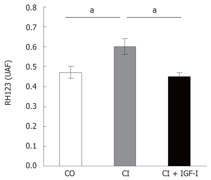
Figure 3 Intramitochondrial ROS production in isolated mitochondria: mitochondria from untreated cirrhotic rats showed an increased production of free radicals as compared to healthy controls (CO) and cirrhotic rats treated with IGF-I (CI + IGF).
mean ± SEM; aP < 0.05.
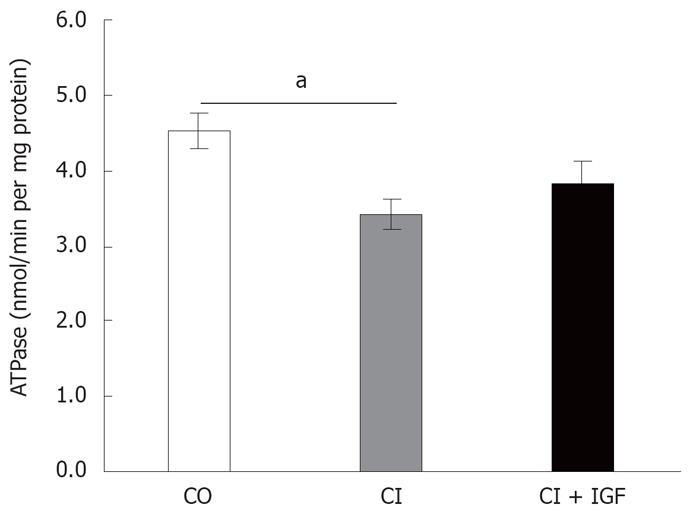
Figure 4 Complex V ATPase activity: ATPase activity was significantly reduced in mitochondria from untreated cirrhotic rats (P < 0.
05 vs CO group). IGF-therapy induced a little increase of ATPase activity (P = no significant vs CO group). mean ± SEM; aP < 0.05.
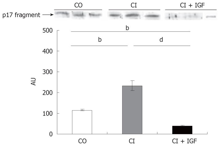
Figure 5 Active fragment of caspase 3 by Western blot in liver homogenates: untreated cirrhotic rats showed an increment of caspase activation.
IGF-I replacement therapy induced an inhibition of caspase 3 activation. bP < 0.01; dP < 0.001.
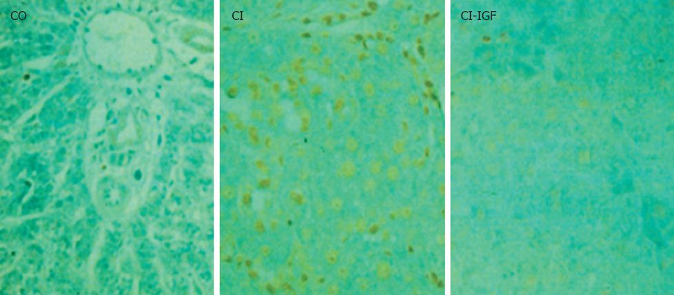
Figure 6 Apoptosis by TUNEL in the liver from the three experimental groups: the number of TUNEL positive hepatocytes was increased in untreated cirrhotic rats (CI) as compared to healthy controls (CO).
IGF-Itherapy reduced signifincantly apoptosis in hepatocytes.
- Citation: Pérez R, García-Fernández M, Díaz-Sánchez M, Puche JE, Delgado G, Conchillo M, Muntané J, Castilla-Cortázar I. Mitochondrial protection by low doses of insulin-like growth factor-Iin experimental cirrhosis. World J Gastroenterol 2008; 14(17): 2731-2739
- URL: https://www.wjgnet.com/1007-9327/full/v14/i17/2731.htm
- DOI: https://dx.doi.org/10.3748/wjg.14.2731


