Copyright
©The Author(s) 2020.
Artif Intell Gastrointest Endosc. Dec 28, 2020; 1(3): 44-52
Published online Dec 28, 2020. doi: 10.37126/aige.v1.i3.44
Published online Dec 28, 2020. doi: 10.37126/aige.v1.i3.44
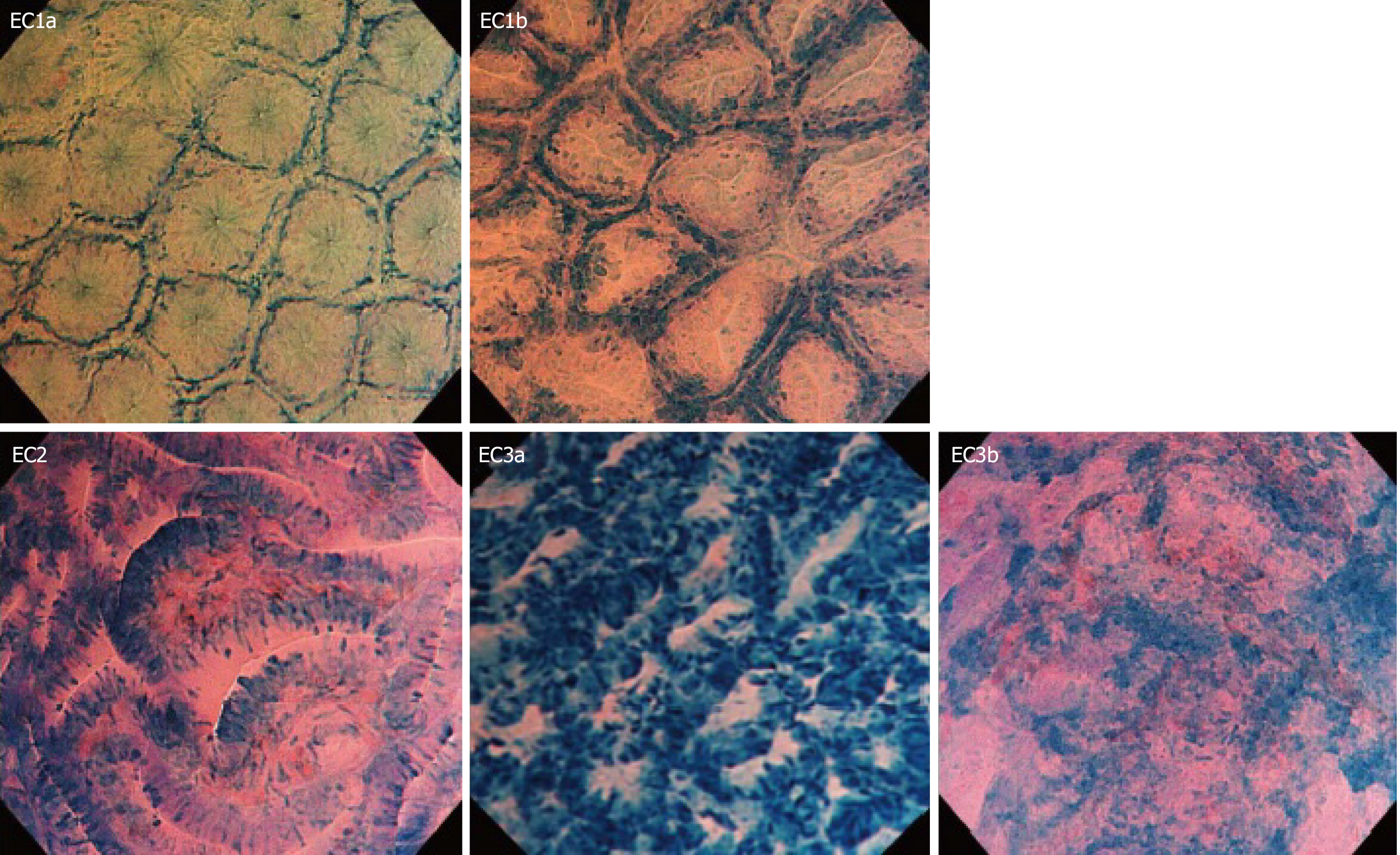
Figure 1 Endocytoscopy classification.
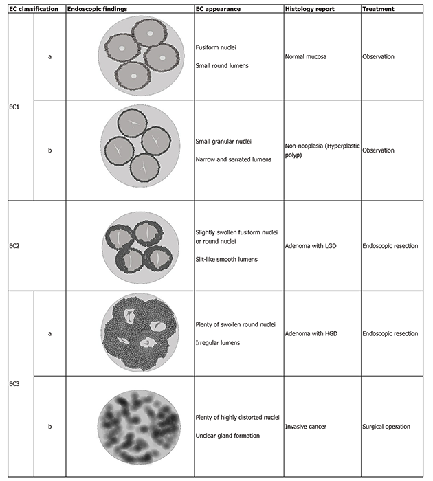
This evaluation system includes endocytoscopy (EC) 1a, fusiform nuclei, and small round gland lumens - normal mucosa; EC1b, small granular nuclei and narrow, serrated gland lumens - non-neoplasia (hyperplastic polyps); EC2, slightly swollen fusiform nuclei or round nuclei and slit-like smooth gland lumens - adenoma with low-grade dysplasia; EC3a, plenty d swollen round nuclei and irregular gland lumens – adenoma with high-grade dysplasia, EC3b, plenty of highly distorted nuclei and unclear gland formation - invasive cancer. EC: endocytoscopy. This figure has been used with the permission of reference[5].
Figure 2 Endocytoscopy classification with endoscopic findings.
EC: Endocytoscopy; LGD: Low-grade dysplasia; HGD: High-grade dysplasia.
- Citation: Peshevska-Sekulovska M, Velikova TV, Peruhova M. Artificial intelligence assisted endocytoscopy: A novel eye in endoscopy. Artif Intell Gastrointest Endosc 2020; 1(3): 44-52
- URL: https://www.wjgnet.com/2689-7164/full/v1/i3/44.htm
- DOI: https://dx.doi.org/10.37126/aige.v1.i3.44


