Copyright
©The Author(s) 2020.
Artif Intell Cancer. Nov 28, 2020; 1(4): 51-65
Published online Nov 28, 2020. doi: 10.35713/aic.v1.i4.51
Published online Nov 28, 2020. doi: 10.35713/aic.v1.i4.51
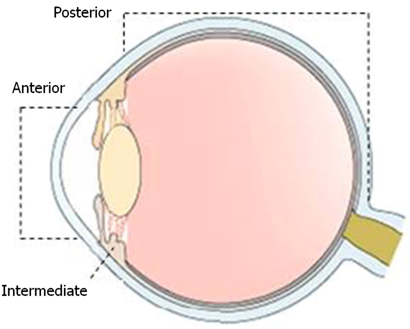
Figure 1 Diagram of sagittal section of the eye.
Portions of the uvea: Anterior (iris), intermediate (ciliary body), and posterior (choroid).
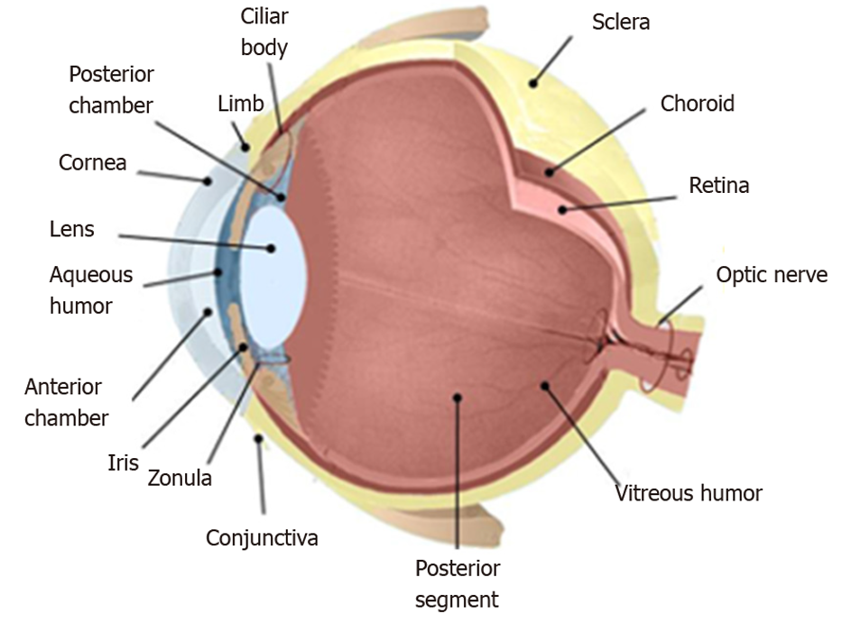
Figure 2 Anatomy of the eye: Eyeball, tunics and layers of the eye, and intraocular structures.
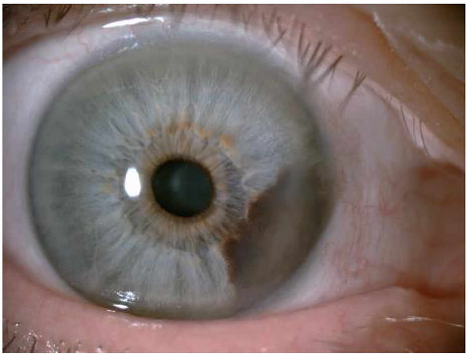
Figure 3 Iris melanoma.
Iris melanic neoformation that extends to the ciliary body.
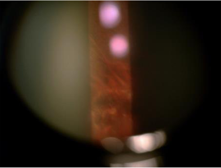
Figure 4 Choroidal nevus in a patient with choroidal melanoma of the contralateral eye.
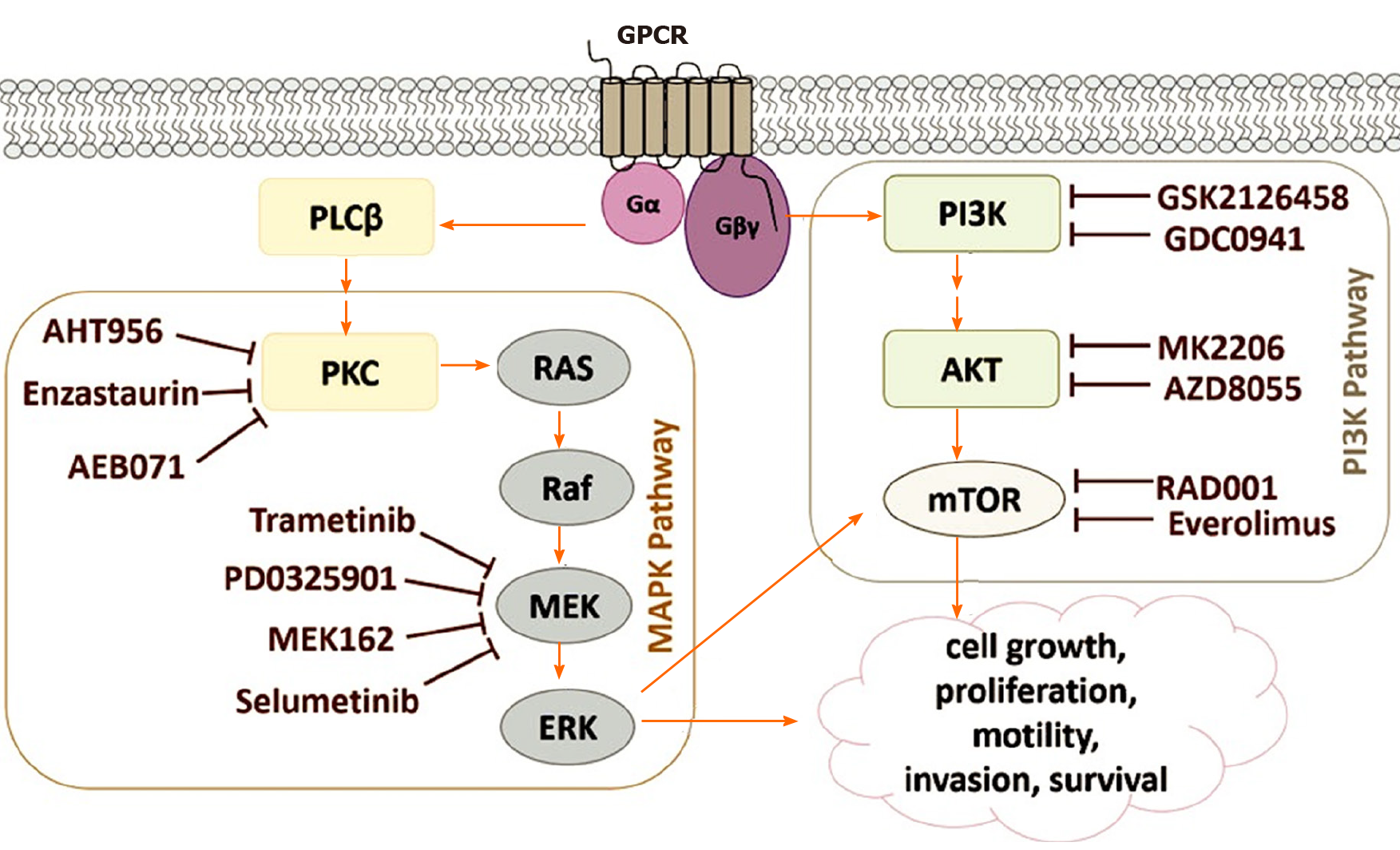
Figure 5 Signaling pathways involved in the development of uveal melanoma.
Figure 6 Schematic representation of a dummy cell.
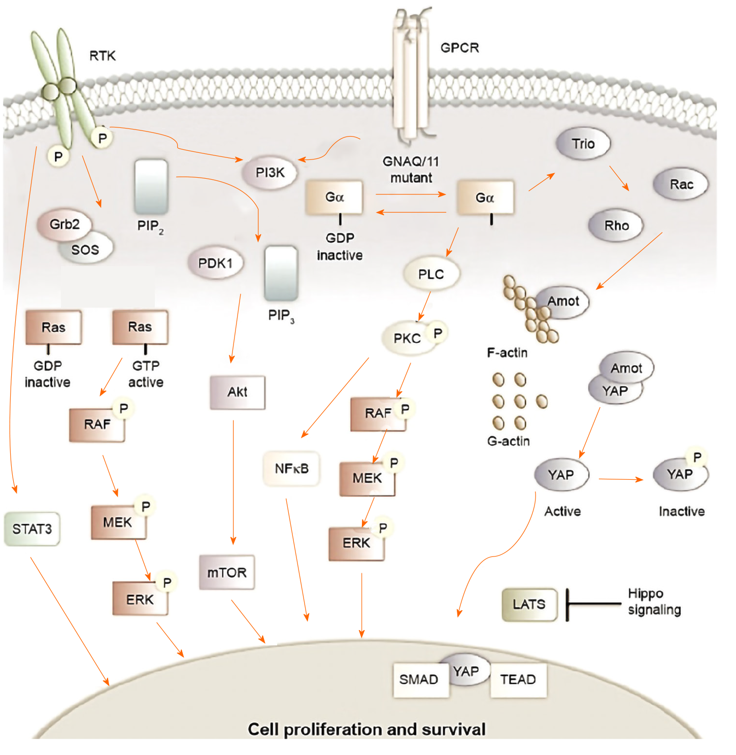
Figure 7 Signaling pathways in uveal melanoma.
Figure 8 Outline of the 3820c.
PIP3.from.PIP2 rewrite rule.
- Citation: Santos-Buitrago B, Santos-García G, Hernández-Galilea E. Artificial intelligence for modeling uveal melanoma. Artif Intell Cancer 2020; 1(4): 51-65
- URL: https://www.wjgnet.com/2644-3228/full/v1/i4/51.htm
- DOI: https://dx.doi.org/10.35713/aic.v1.i4.51


