Copyright
©The Author(s) 2019.
World J Meta-Anal. May 31, 2019; 7(5): 218-223
Published online May 31, 2019. doi: 10.13105/wjma.v7.i5.218
Published online May 31, 2019. doi: 10.13105/wjma.v7.i5.218
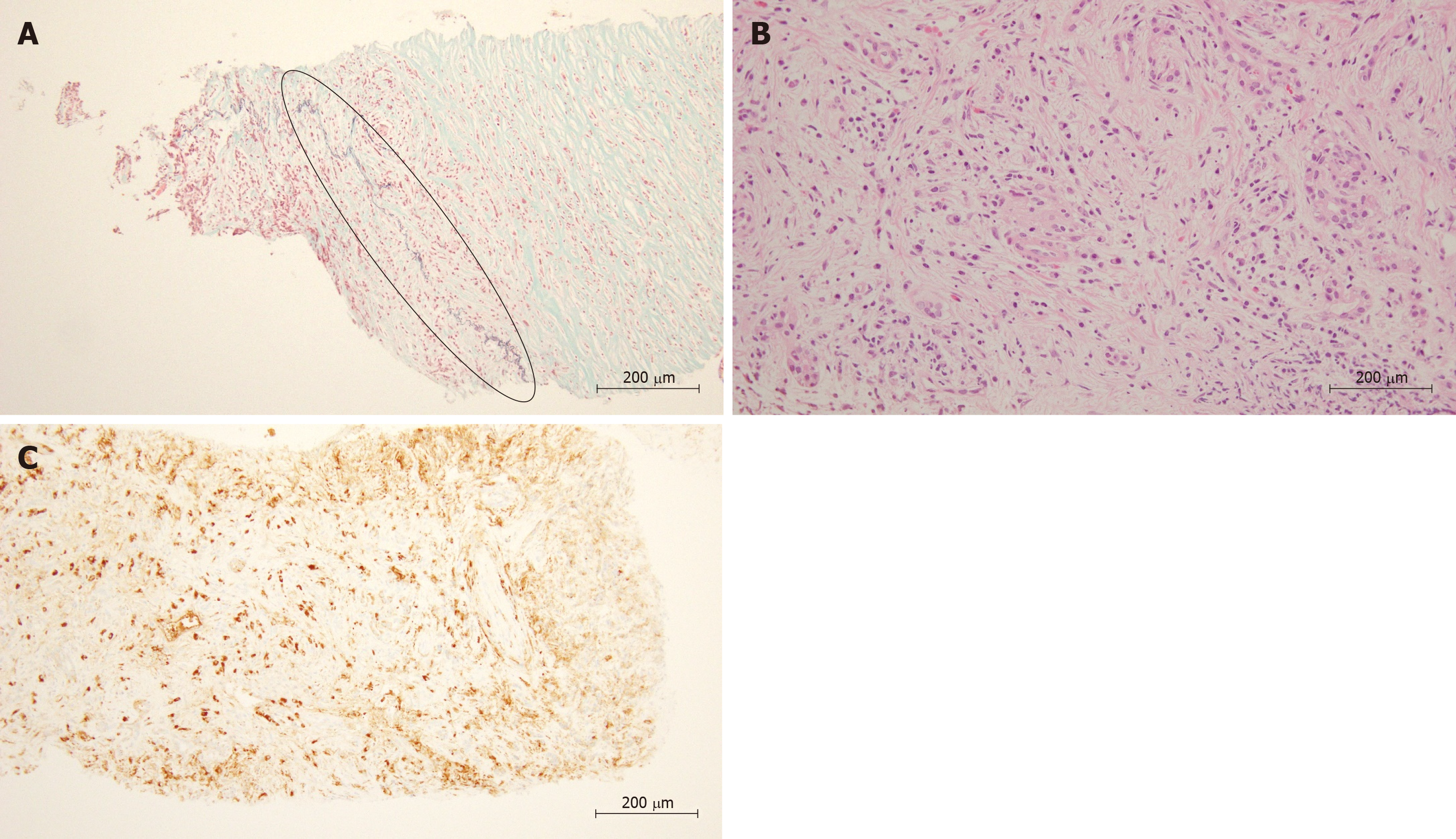
Figure 1 Histological findings in lymphoplasmacytic sclerosing pancreatitis by endoscopic ultrasonography-guided fine needle aspiration.
A: Obliterative phlebitis was observed, highlighted in an ellipse (EM × 200); B: Plasma cells and storiform fibrosis were observed (HE × 400); C: IgG4-positive plasma cells were observed (IgG4 immunostaining × 200).
- Citation: Sugimoto M, Takagi T, Suzuki R, Konno N, Asama H, Sato Y, Irie H, Watanabe K, Nakamura J, Kikuchi H, Takasumi M, Hashimoto M, Hikichi T, Ohira H. Present state of endoscopic ultrasonography-guided fine needle aspiration for the diagnosis of autoimmune pancreatitis type 1. World J Meta-Anal 2019; 7(5): 218-223
- URL: https://www.wjgnet.com/2308-3840/full/v7/i5/218.htm
- DOI: https://dx.doi.org/10.13105/wjma.v7.i5.218


