Copyright
©The Author(s) 2024.
World J Meta-Anal. Sep 18, 2024; 12(3): 94519
Published online Sep 18, 2024. doi: 10.13105/wjma.v12.i3.94519
Published online Sep 18, 2024. doi: 10.13105/wjma.v12.i3.94519
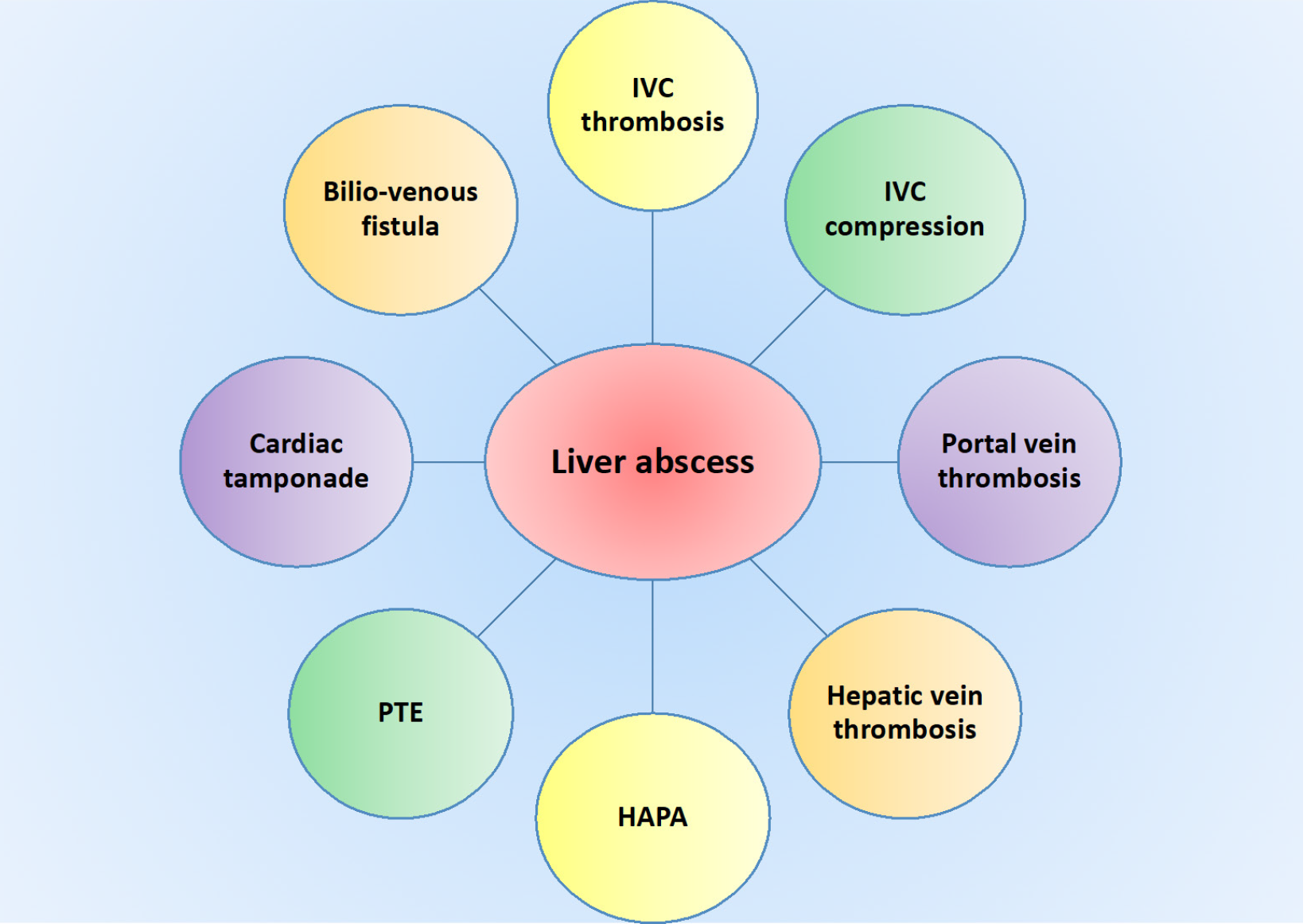
Figure 1 Spectrum of vascular complications associated with liver abscess.
IVC: Inferior vena cava; HAPA: Hepatic artery pseudoaneurysm; PTE: Pulmonary thromboembolism.
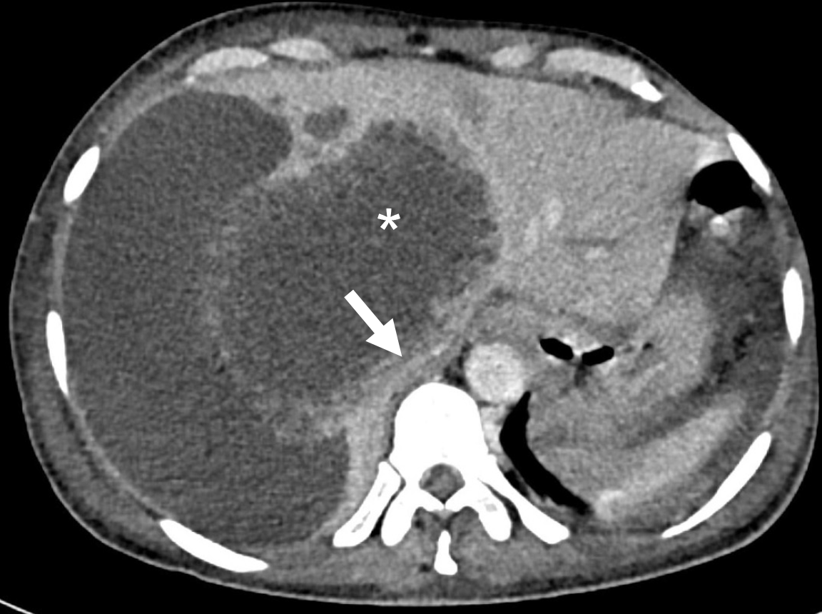
Figure 2 Axial contrast-enhanced computed tomography image showing a large ruptured amebic liver abscess (asterisk) in the caudate lobe, exerting pressure on the inferior vena cava (arrow).
Note the fluid collection in perihepatic space resulting from the ruptured abscess.
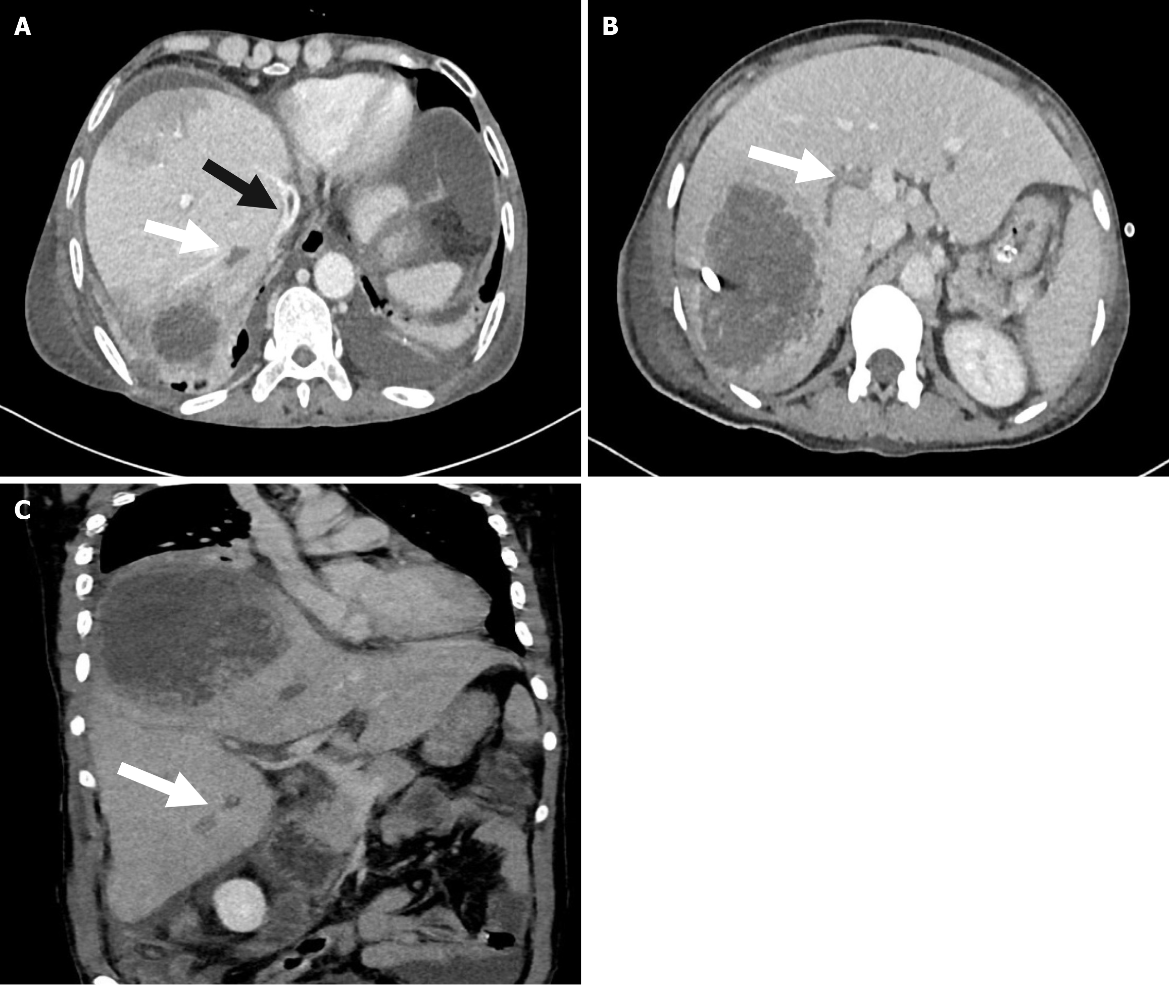
Figure 3 Venous thrombosis in associations with liver abscess.
A: Axial contrast-enhanced computed tomography (CT) imaging illustrates the presence of thrombosis within the inferior vena cava (black arrow) and the right hepatic vein (white arrow) in a patient with abscess in right lobe of liver; B: Axial contrast-enhanced CT scan demonstrates the presence of a thrombus within the right posterior segmental branch of the right portal vein in a patient with liver abscess; C: Coronal contrast-enhanced CT imaging depicts thrombosis occurring in the segmental branch of the right portal vein in a patient with liver abscess.
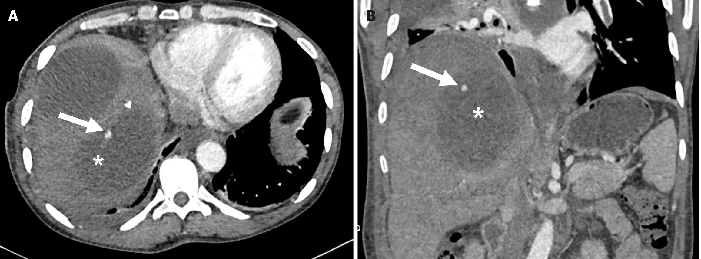
Figure 4 Computed tomography scan image of hepatic artery pseudoaneurysm in association with liver abscess.
A: Axial image demonstrating a small aneurysm (arrow) originating from a peripheral branch of the right hepatic artery within the abscess cavity (asterisk); B: Coronal computed tomography image of the same patient demonstrating the hepatic artery pseudoaneurysm.
- Citation: Arya R, Kumar R, Priyadarshi RN, Narayan R, Anand U. Vascular complications of liver abscess: A literature review. World J Meta-Anal 2024; 12(3): 94519
- URL: https://www.wjgnet.com/2308-3840/full/v12/i3/94519.htm
- DOI: https://dx.doi.org/10.13105/wjma.v12.i3.94519


