Copyright
©The Author(s) 2021.
World J Clin Cases. Jun 6, 2021; 9(16): 3908-3913
Published online Jun 6, 2021. doi: 10.12998/wjcc.v9.i16.3908
Published online Jun 6, 2021. doi: 10.12998/wjcc.v9.i16.3908
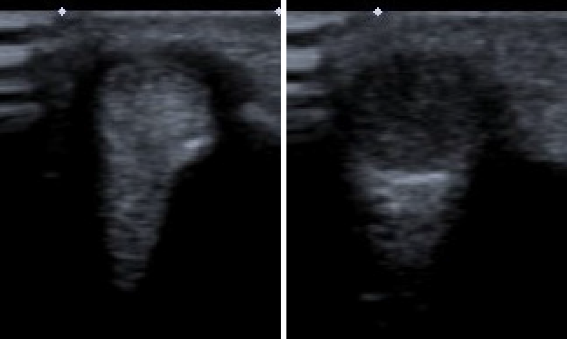
Figure 1 Dynamic sonography showed tendon jumping when wrist snapping.
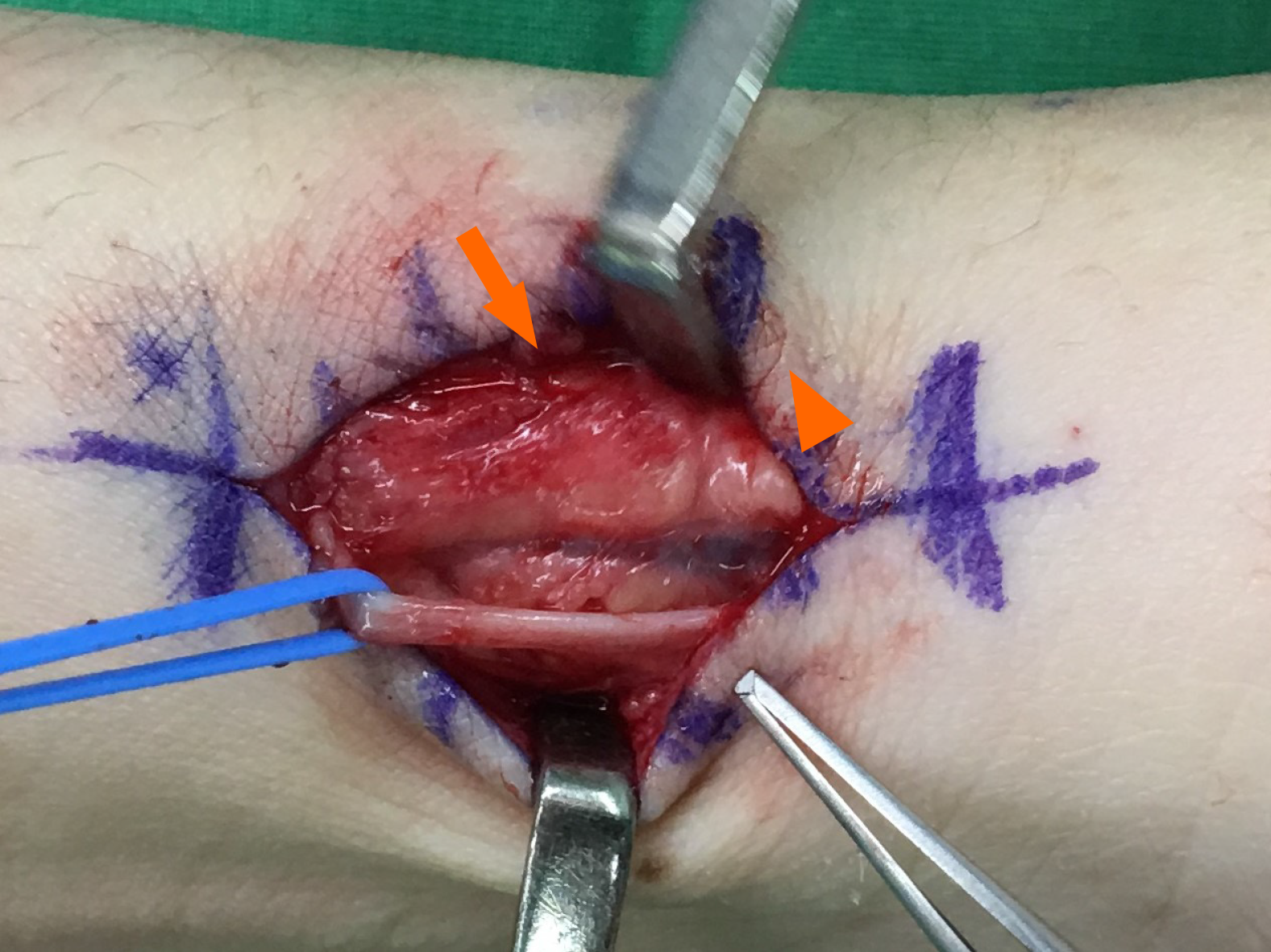
Figure 2 Tendon gliding out of the distal extensor retinaculum (arrow), forming a soft tissue ball (arrow head).
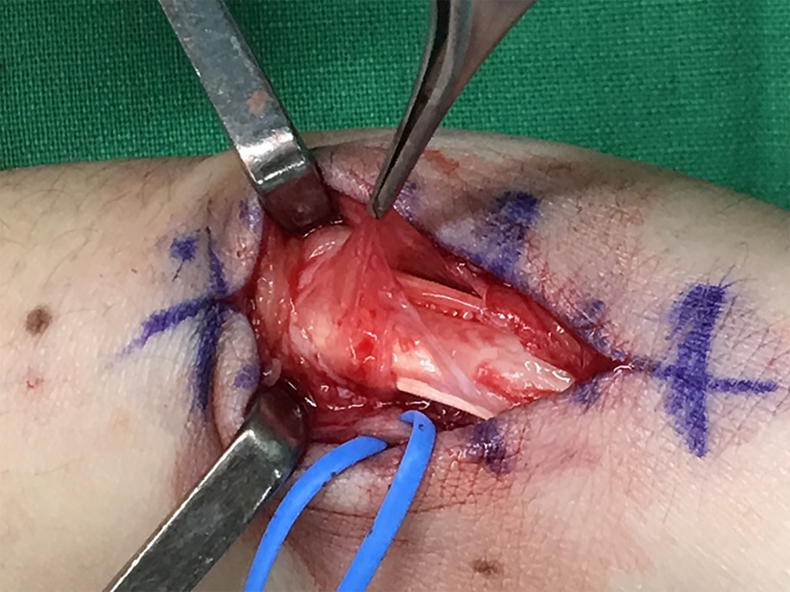
Figure 3 The soft tissue ball was comprised of proliferated synovium from two slips of abductor pollicis longus.
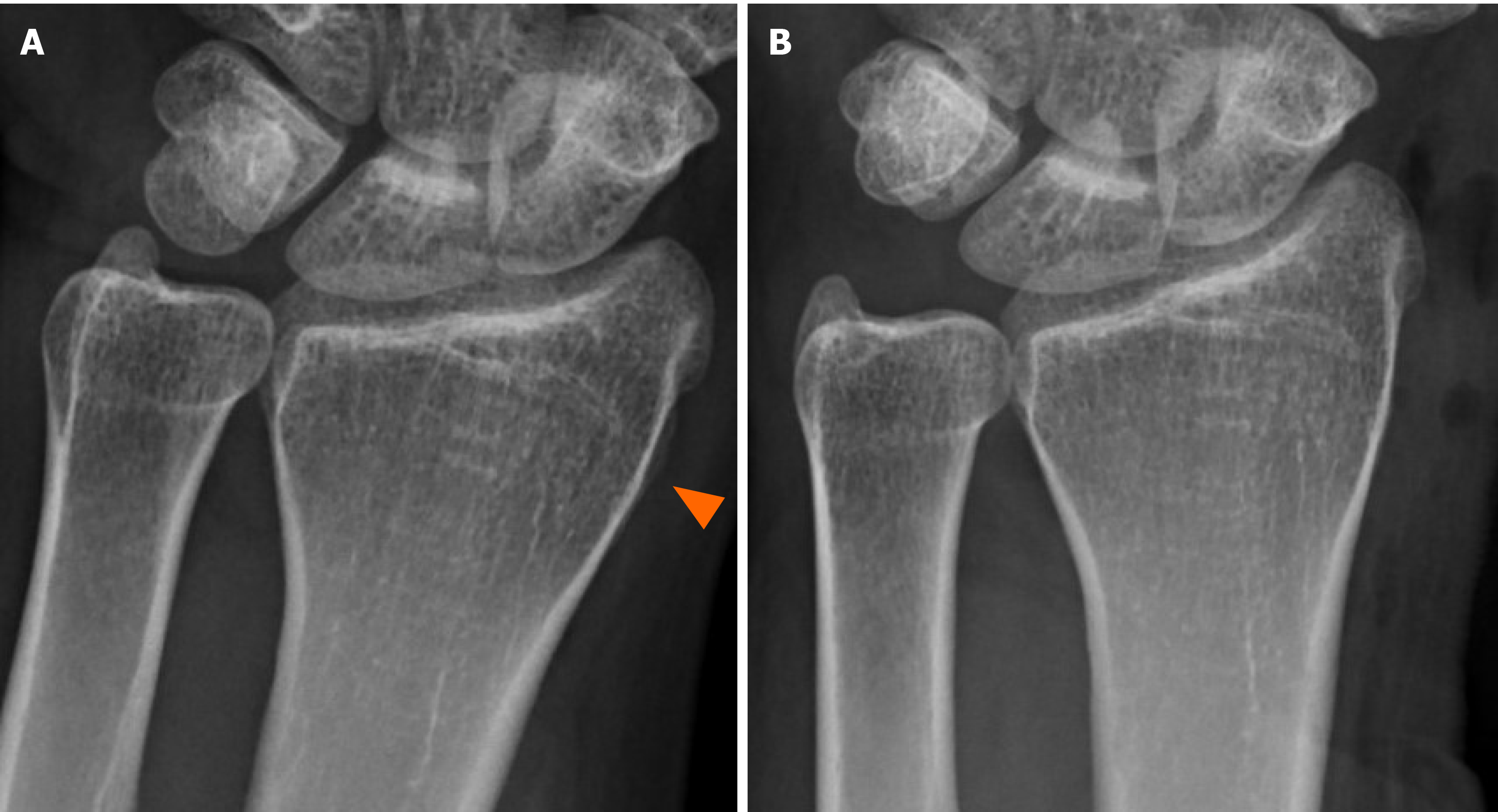
Figure 4 Radiography.
A: Overgrown bone (arrow head) before excision; B: Overgrown bone after excision.
- Citation: Hu CJ, Chow PC, Tzeng IS. Snapping wrist due to bony prominence and tenosynovitis of the first extensor compartment: A case report. World J Clin Cases 2021; 9(16): 3908-3913
- URL: https://www.wjgnet.com/2307-8960/full/v9/i16/3908.htm
- DOI: https://dx.doi.org/10.12998/wjcc.v9.i16.3908


