Copyright
©2013 Baishideng Publishing Group Co.
World J Methodol. Sep 26, 2013; 3(3): 27-38
Published online Sep 26, 2013. doi: 10.5662/wjm.v3.i3.27
Published online Sep 26, 2013. doi: 10.5662/wjm.v3.i3.27
Figure 1 Flow chart of experimental protocol.
cMRI: Cardiac magnetic resonance imaging; DE: Delayed enhancement; RIO: Red-iodized-oil; DR: Digital radiography; HE: Hematoxylin-eosin.
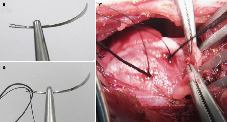
Figure 2 Double suture method in rabbit model of reperfused myocardial infarction.
A: A 1/2 circle shape triangular needle has spring eyes at the end; B: Two silk sutures were placed through separate eyes; C: The thicker suture (2-0) was used for the left circumflex coronary artery (LCx) ligation, which would be removed for reperfusion, and the thinner one (5-0) was spared for later ex vivo LCx re-occlusion in order to perform postmortem bifunctional staining.
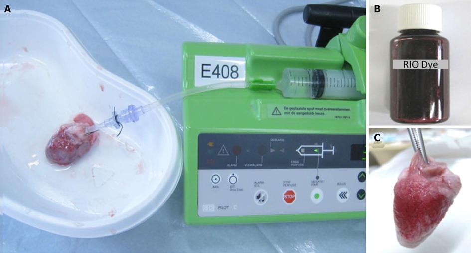
Figure 3 Bifunctional staining methods.
A: A plastic catheter filled with saline was inserted into the aorta and anchored with its tip 1 cm above the aortic valves, which was connected to a 10 mL syringe fixed in the injection bump. The heart was gently rinsed to wash out remaining blood, then the left circumflex coronary artery was re-ligated, and 2-4 mL of red iodized oil (RIO) dye was infused; B: Homemade RIO dye; C: Lateral view of the RIO perfused heart. The perfusion of RIO dye was stopped when the normal myocardium was stained completely scarlet red, while the area at risk remained unstained.
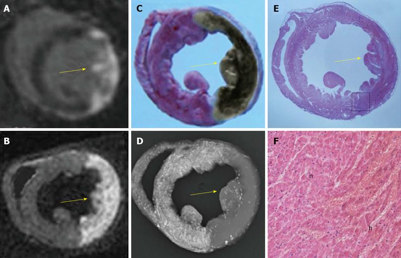
Figure 4 An example of a reperfused hemorrhagic myocardial infarction (arrow) shown by cardiac magnetic resonance imaging and the area at risk demonstrated with bifunctional staining method.
A: The hyperenhanced transmural myocardial infarction (MI) was seen at the lateral wall on in vivo delay enhancement (DE) cardiac magnetic resonance imaging (cMRI); B: The ex vivo DE cMRI was highly correspondent with A; C: In contrast to the normal myocardium with red staining, the area at risk (AAR) was shown as an unstained region, on which the brown grayish area indicates a large intramural hemorrhagic infarction after the fixation by Formalin; D: On digital radiography (DR), the AAR was shown as non-opacified region in contrast to the opacified normal myocardium. Notice that the AAR defined by red iodized oil-staining (RIO-staining) in C and DR in D was somewhat larger than the MI defined by in vivo DE cMRI in A and ex vivo DE cMRI in B; E: The hematoxylin-eosin (HE) stained macroscopic view was obtained from the corresponding histological section of A, B, C, and D; F: Photomicroscopic view of the HE stained slice (magnification, × 100) confirmed the presence of myocardial necrosis with hemorrhage and tissue reaction. h: Hemorrhagic infarction; n: Adjacent normal myocardium with inflammatory infiltration.
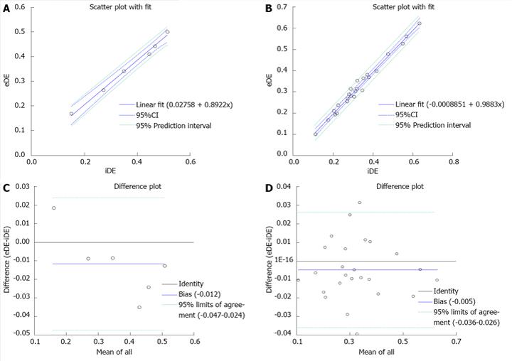
Figure 5 The linear correlation (above) and Bland-Altman analyses (bottom) plots comparing the myocardial infarction on in vivo and ex vivo delayed enhancement magnetic resonance imaging.
The plots graph displayed a good correlation between MI measured with in vivo delayed enhancement (iDE) and ex vivo delayed enhancement (eDE) on global (A) and slices (B). Plots of differences in myocardial infarction on global (C) and slices (D) were indicative of high agreement between iDE and eDE.
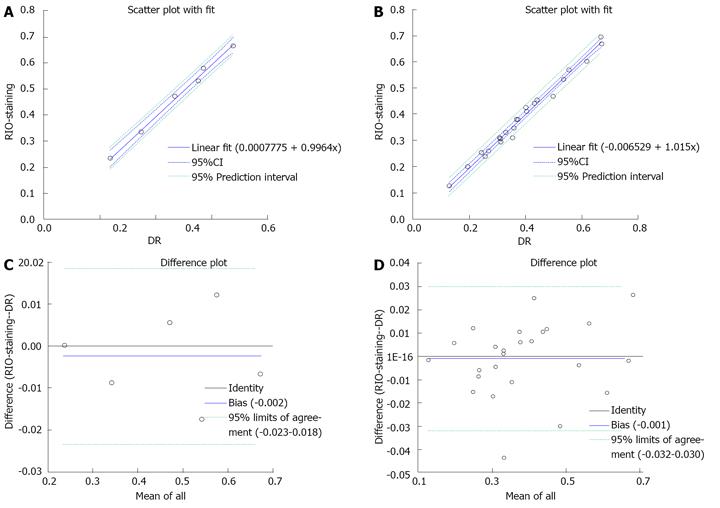
Figure 6 The linear correlations (above) and Bland-Altman analyses (bottom) to compare the area at risk by red iodized oil-staining and digital radiography of the bifunctional staining.
The plots showed a good correlation between the area at risk (AAR) measured with red iodized oil (RIO)-staining and digital radiography (DR) on global (A) and slices (B). Plots of the differences in AAR on global (C) and slices (D) were almost zero, indicating that RIO-staining and DR are essentially equivalent.
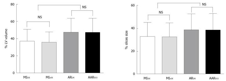
Figure 7 Comparisons between myocardial infarction and area at risk by different techniques.
There were no significant differences between myocardial infarction in vivo delayed enhancement (MI iDE) and myocardial infarction ex vivo delayed enhancement (MI eDE) measured from cardiac magnetic resonance imaging in global (left) and slices (right). There were no significant differences between area at risk digital radiography (AARDR) and area at risk red iodized oil-staining (AARRIO-staining) in global (left) and slices (right).The significant differences were observed on AARDRvs MI iDE (P < 0.01); AARDRvs MI eDE (P < 0.01); AARRIO-stainingvs MI iDE (P < 0.01); AARRIO-stainingvs MI eDE (P < 0.01). NS: Not significant.
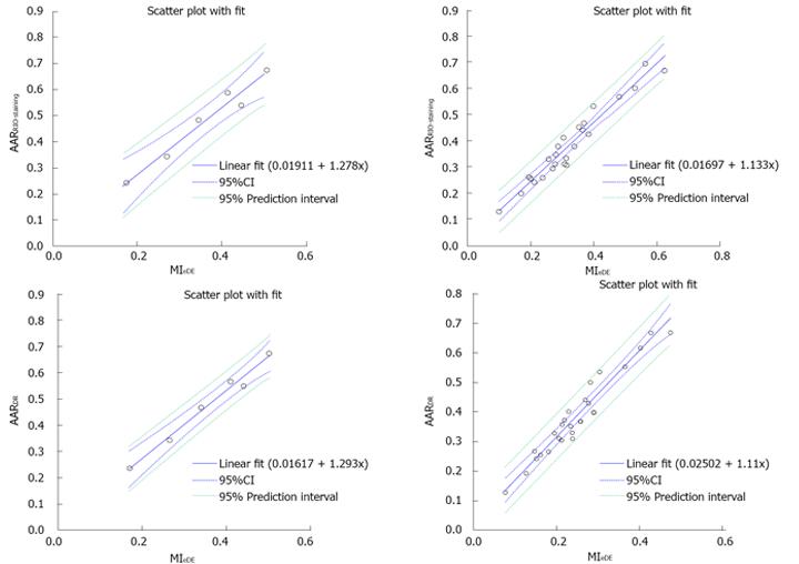
Figure 8 The linear correlations between ex vivo cardiac magnetic resonance imaging and bifunctional staining.
The myocardial infarction (MI) measured by global (left) and slices (right) on ex vivo delayed enhancement cardiac magnetic resonance imaging corresponded well with area at risk by red iodized oil-staining (above) and digital radiography (bottom), confirming the bifunctional staining with RIO dye could successfully delineate and characterize the AAR. MIeDE: Myocardial infarction ex vivo delayed enhancement; AARRIO-staining: Area at risk on red-iodized oil-staining; AARDR: Area at risk on digital radiography.
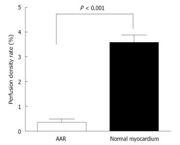
Figure 9 Comparison of perfusion density rate between area at risk and normal myocardium.
Coronary artery occlusion caused a significant decrease of blood perfusion in ischemic region compared to non-ischemic region (P < 0.001), resulted in a difference over 10 times in-between. AAR: Area at risk.
-
Citation: Feng Y, Ma ZL, Chen F, Yu J, Cona MM, Xie Y, Li Y, Ni Y. Bifunctional staining for
ex vivo determination of area at risk in rabbits with reperfused myocardial infarction. World J Methodol 2013; 3(3): 27-38 - URL: https://www.wjgnet.com/2222-0682/full/v3/i3/27.htm
- DOI: https://dx.doi.org/10.5662/wjm.v3.i3.27


