Copyright
©The Author(s) 2025.
World J Methodol. Dec 20, 2025; 15(4): 107411
Published online Dec 20, 2025. doi: 10.5662/wjm.v15.i4.107411
Published online Dec 20, 2025. doi: 10.5662/wjm.v15.i4.107411
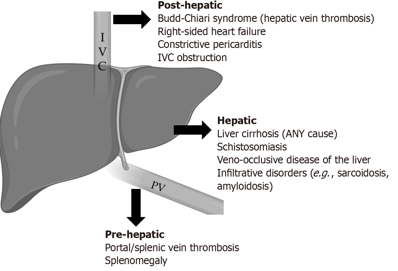
Figure 1 Some causes of portal hypertension.
IVC: Inferior vena cava; PV: Portal vein. Image created using Biorender.
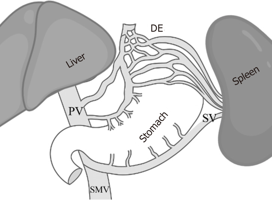
Figure 2 Portal circulation and related anatomy.
The stomach’s venous drainage is into the splenic vein, while the esophageal drainage (especially lower esophagus) is into the portal vein (PV), which is the reason for preferential variceal development at the lower esophagus in cirrhotic portal hypertension (PH), whereas the non-cirrhotic PH there is mainly the development of gastric varices. DE: Distal esophagus; SMV: Superior mesenteric vein; SV: Splenic vein. Image created using Biorender.
- Citation: Abdulrasak M, Ahmed M, Hootak S. Utility of splenic transient elastography in assessing the presence of portal hypertension: A review. World J Methodol 2025; 15(4): 107411
- URL: https://www.wjgnet.com/2222-0682/full/v15/i4/107411.htm
- DOI: https://dx.doi.org/10.5662/wjm.v15.i4.107411


