Copyright
©The Author(s) 2023.
World J Methodol. Dec 20, 2023; 13(5): 502-509
Published online Dec 20, 2023. doi: 10.5662/wjm.v13.i5.502
Published online Dec 20, 2023. doi: 10.5662/wjm.v13.i5.502
Figure 1 X-ray.
A: Antero-posterior preoperative X-ray of both hips where severe bilateral osteoarthritis is observed. A marked narrow endomedular canal is observed; B: Anteroposterior X-ray of both hips 5 years after operation, showing rupture at the proximal middle third level of the femoral stem; C: Immediate postoperative X-ray, showing femoral revision surgery. Trochanteric osteotomy and a poor proximal cement mantle are shown; D: Anteroposterior X-ray of both hips, 2 years after revision surgery, showing a further rupture of the Exeter® stem, at the level of prior femoral osteotomy.
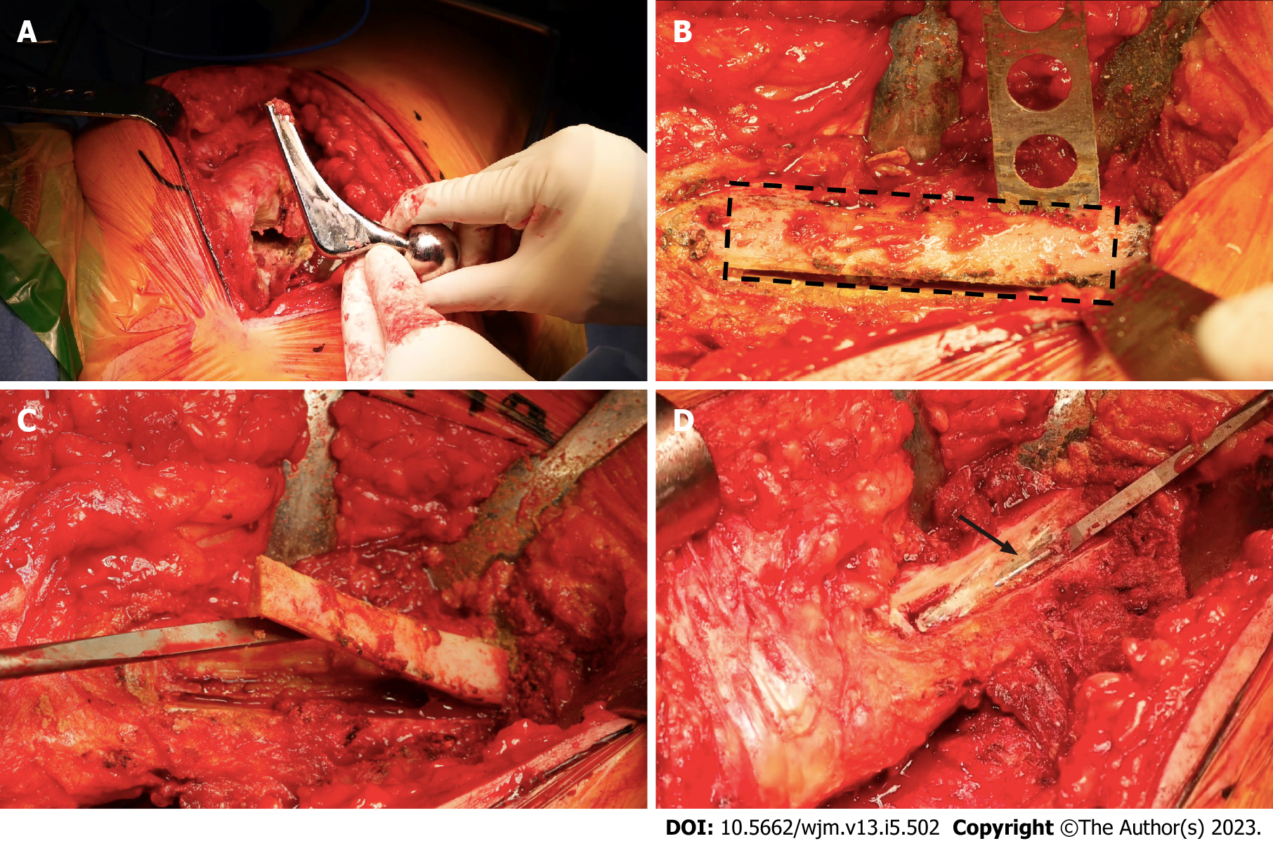
Figure 2 Surgical operation.
A: Intraoperative images of implant removal during re-revision surgery, showing the removal of the proximal part of the broken femoral stem; B: Planning and execution of lateral or window femoral osteotomy (black dotted line) with oscillating saw; C: Osteotomized segment of the window and exposure of the medullary canal; D: Removing the cement and distal segment of the stem (black arrow).
Figure 3 Femoral broaching was performed in order to achieve good contact between the host bone and the distal fixation stem.
A: Intraoperative images during re-revision surgery. Definitive cementless distal fixation modular stem; B: Direct view of the insertion of the new stem, through the femoral window; C: Osteosynthesis of the osteotomized segment with wires; D: A femur Anteroposterior X-ray, where the final immediate postoperative result is shown with an uncemented distal fixation femoral stem and osteosynthesis of the cortical window.
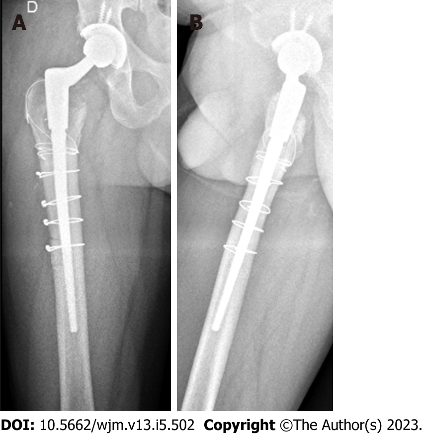
Figure 4 Antero-posterior and lateral X-ray.
A: Lateral X-ray; B: Antero-posterior X-ray. Antero-posterior and lateral X-ray 3 mo after re-revision surgery, in which the correct position of the femoral implant and consolidation of osteotomy are shown.
- Citation: Lucero CM, Luco JB, Garcia-Mansilla A, Slullitel PA, Zanotti G, Comba F, Buttaro MA. Successful hip revision surgery following refracture of a modern femoral stem using a cortical window osteotomy technique: A case report and review of literature. World J Methodol 2023; 13(5): 502-509
- URL: https://www.wjgnet.com/2222-0682/full/v13/i5/502.htm
- DOI: https://dx.doi.org/10.5662/wjm.v13.i5.502


