Copyright
©The Author(s) 2021.
World J Methodol. Jul 20, 2021; 11(4): 208-221
Published online Jul 20, 2021. doi: 10.5662/wjm.v11.i4.208
Published online Jul 20, 2021. doi: 10.5662/wjm.v11.i4.208
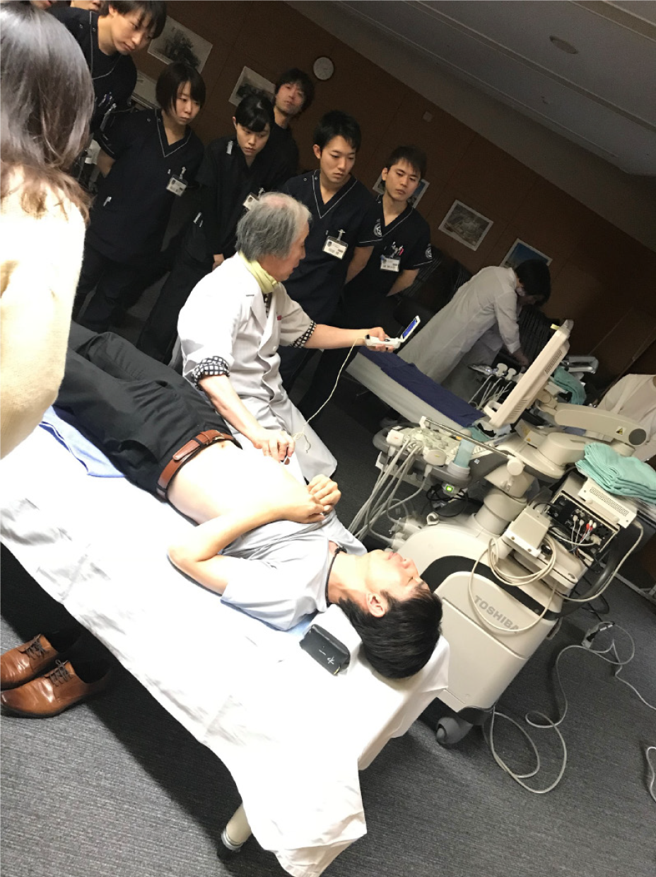
Figure 1 Lecture by a clinician who specializes in ultrasound.
The lecture covers basic ultrasound physics and simple ultrasound anatomy of the abdomen.
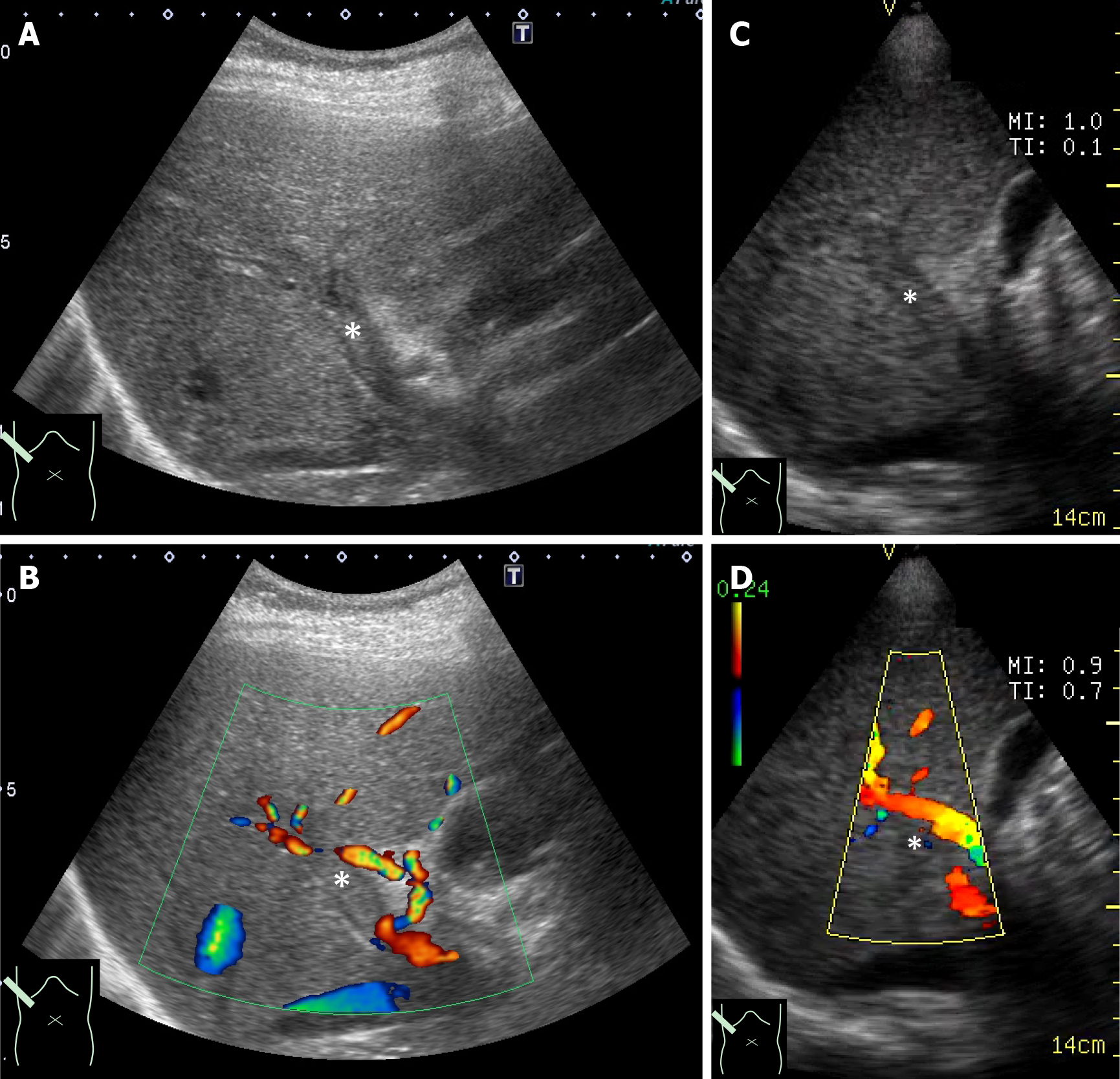
Figure 2 Simultaneous presentation of ultrasound images of a portal thrombus.
A: High-end ultrasound B mode; B: High-end ultrasound color Doppler; C: Pocket-sized ultrasound B mode; D: Pocket-sized ultrasound color Doppler. These comparisons are used to understand the difference in image quality between the two machines. It also confirms that pocket-sized ultrasound is sufficient for diagnoses. *: Thrombus in the portal vein.
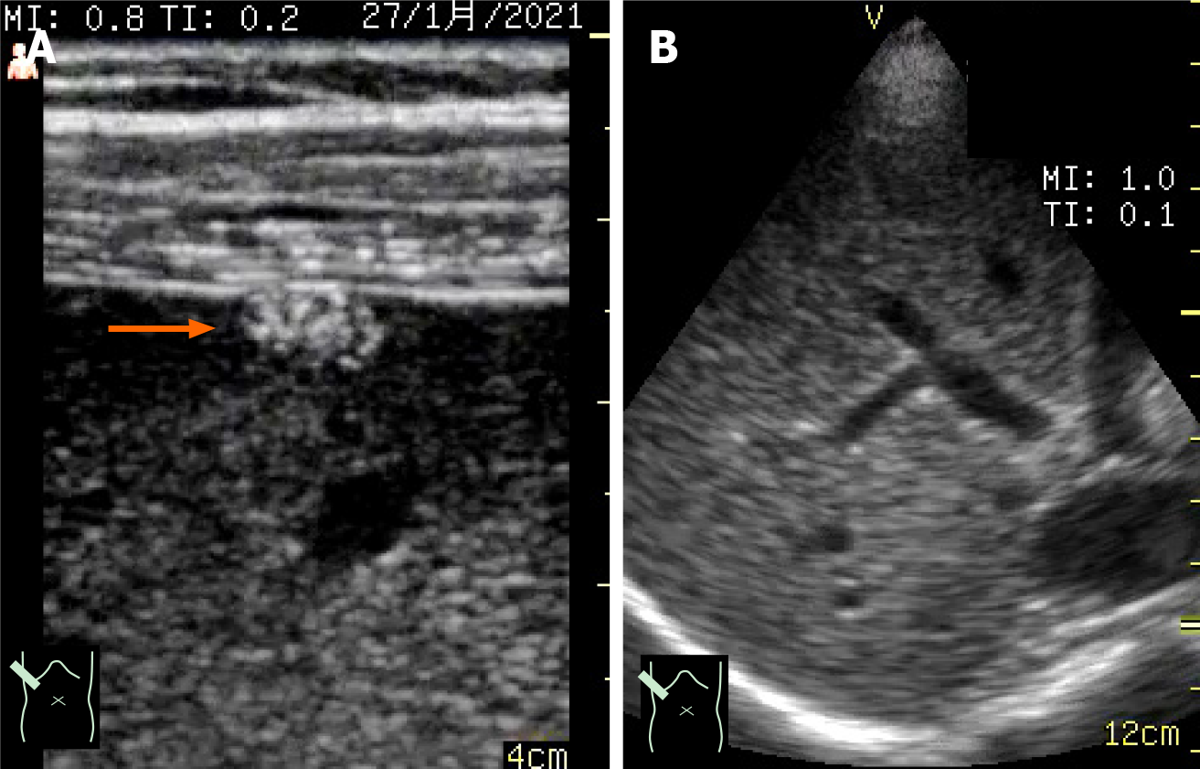
Figure 3 Ultrasound image by different probes on a pocket-sized ultrasound device.
A: High-frequency linear probe; B: 1.7-3.8 MHz sector probe. The linear probe is used to visualize superficial areas, and the sector probe is used for observing deep areas. In this case of a small liver tumor situated at the hepatic surface, the lesion was detected by the linear probe (→) but not the sector probe.
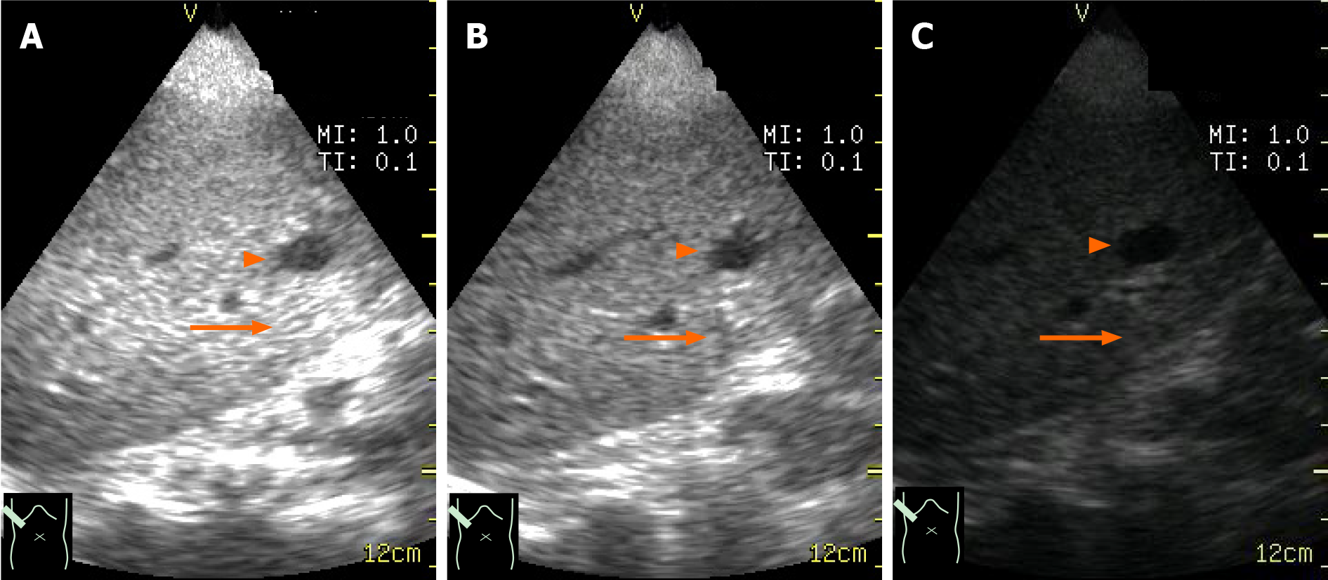
Figure 4 B mode gain setting (hepatic cyst).
A: B mode is too high; B: B mode is well-adjusted; C: B mode is too low. The lesion anechoic mass (arrowhead) with acoustic enhancement (arrow) is clearly seen only when the B mode gain is well-adjusted.
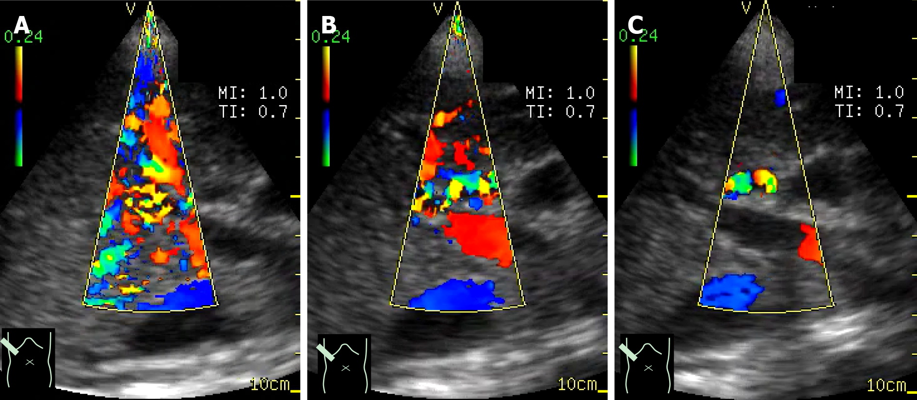
Figure 5 Color Doppler gain setting (normal portal vein).
A: Gain setting is too high; B: Gain setting is well-adjusted; C: Gain setting is too low. Portal venous flow is not detected when color gain setting is too low (C) and is covered by color noise when it is too high (A).
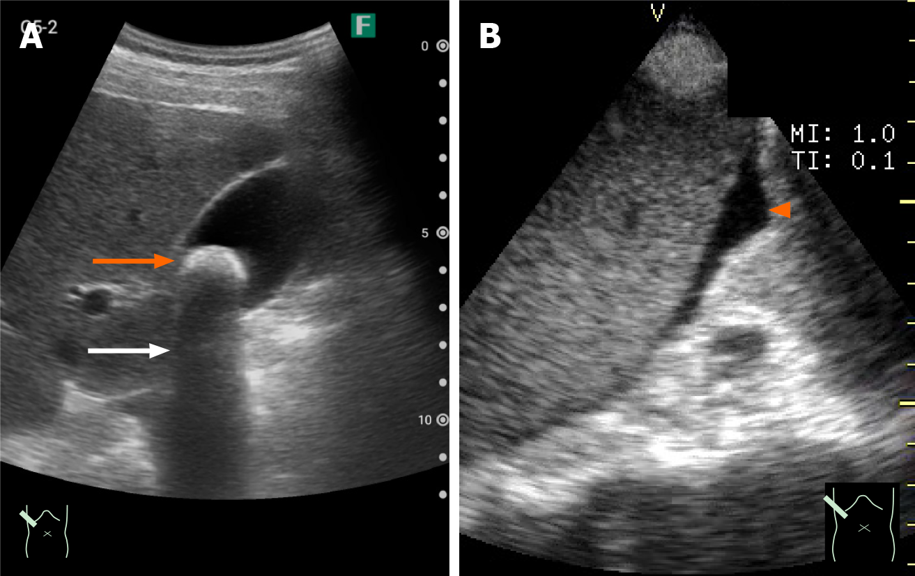
Figure 6 Pocket-sized ultrasound images of gallbladder stones and ascites.
A: A 1-cm stone; B: A small amount of ascites. A 1-cm stone (A) and a small amount of ascites (B) is clearly visualized as a strong echo (orange arrow) with acoustic shadowing (white arrow) in the former and an echo-free space in the latter (arrowhead).
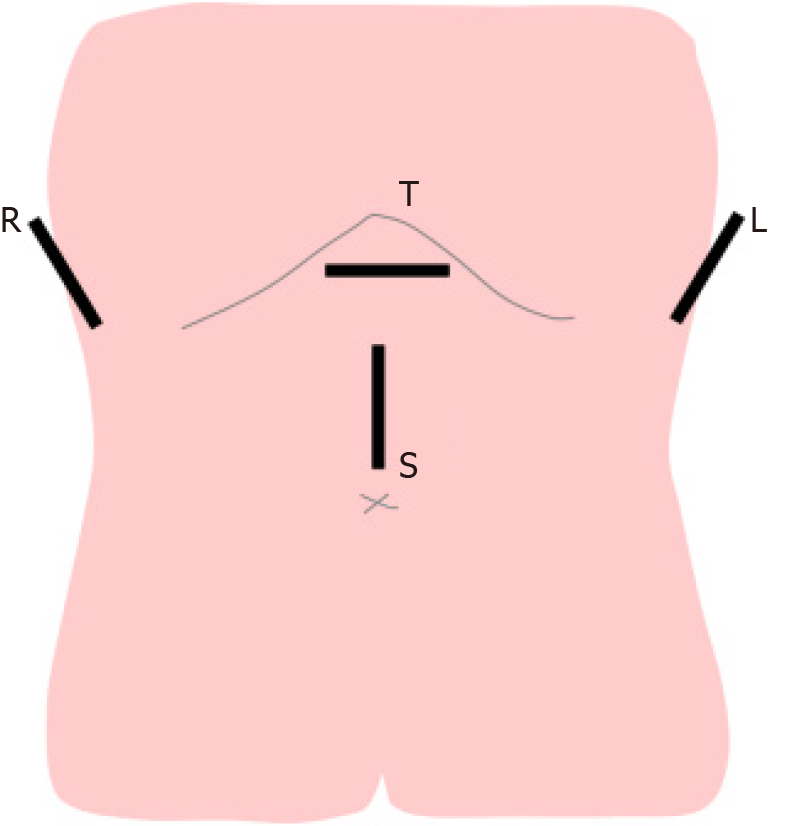
Figure 7 Schematic drawing of basic scanning planes of the abdomen.
The combination of these indicated planes permits quick observation of the whole abdomen. The transverse plane of the upper abdomen is for observation of the pancreas. The right upper abdominal plane is for observation of the liver and the gallbladder. The left upper abdominal plane is for observation of the spleen and left kidney. The sagittal plane is for observation of the abdominal aorta and its branches. L: Left upper plane; R: Right upper plane; S: Sagittal plane; T: Transverse plane.
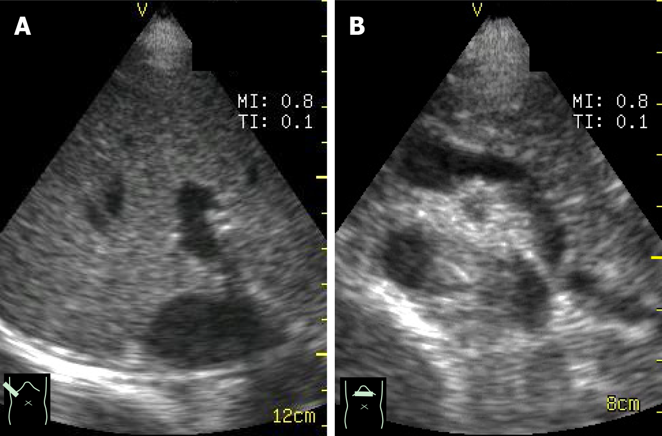
Figure 8 Representative ultrasound images of the abdominal scanning procedure.
A: The right upper abdominal scanning plane permits the observation of the liver (right lobe); B: Through the transverse scanning plane, the pancreas and the neighboring vessels are clearly demonstrated.
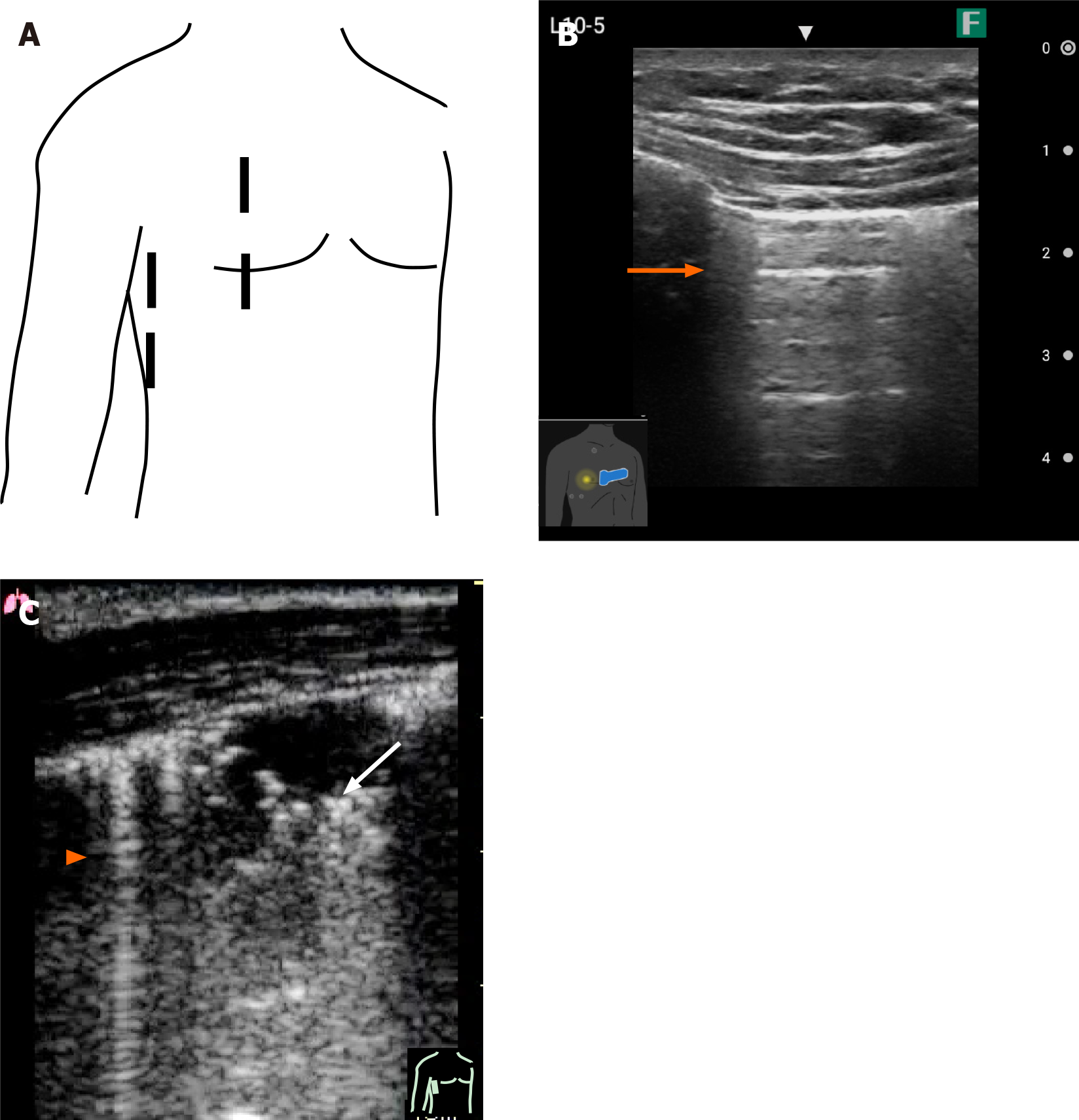
Figure 9 Pocket-sized ultrasound image of the pleura.
A: Basic scanning planes of the chest (right) by ultrasound; B: Ultrasound image of normal pleura showing a typical A line (orange arrow); C: Ultrasound image of B line (arrowhead) and consolidation of the lung (white arrow).
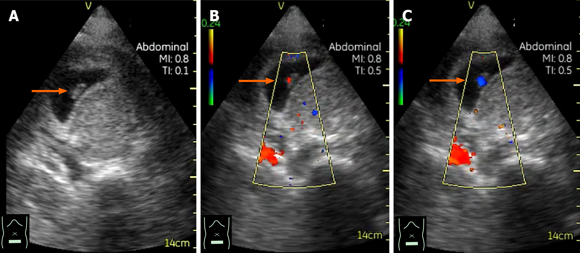
Figure 10 Case presentation 1 of undesirable pregnancy causing abdominal symptoms.
A: At presentation, the patient denied the possibility of pregnancy. However, pocket-sized B mode performed as part of a general physical examination revealed an embryo (arrow) in her lower abdomen. B and C: Color Doppler was particularly useful for confirming the baby’s cardiac movement. Daily use of pocket-sized ultrasound can lead to a reduction in ionizing radiation and time to correct diagnosis.
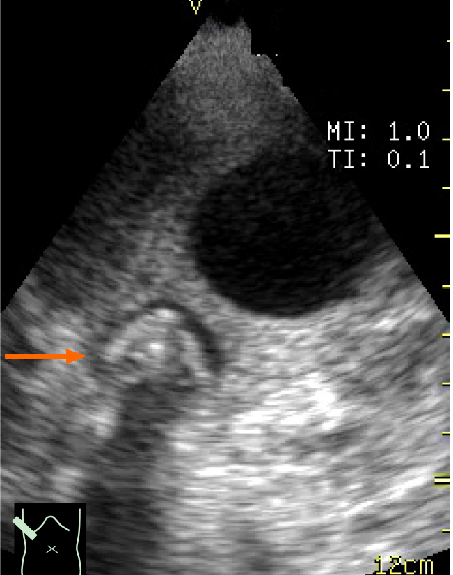
Figure 11 Case presentation 2 of abdominal pain so severe that the patient could not move.
The patient’s abdominal pain was so severe that she could not move from the ambulance. Pocket-sized ultrasound performed in the ambulance revealed a gallbladder stone impact (arrow), leading to the diagnosis of acute stone-impact-induced cholecystitis.
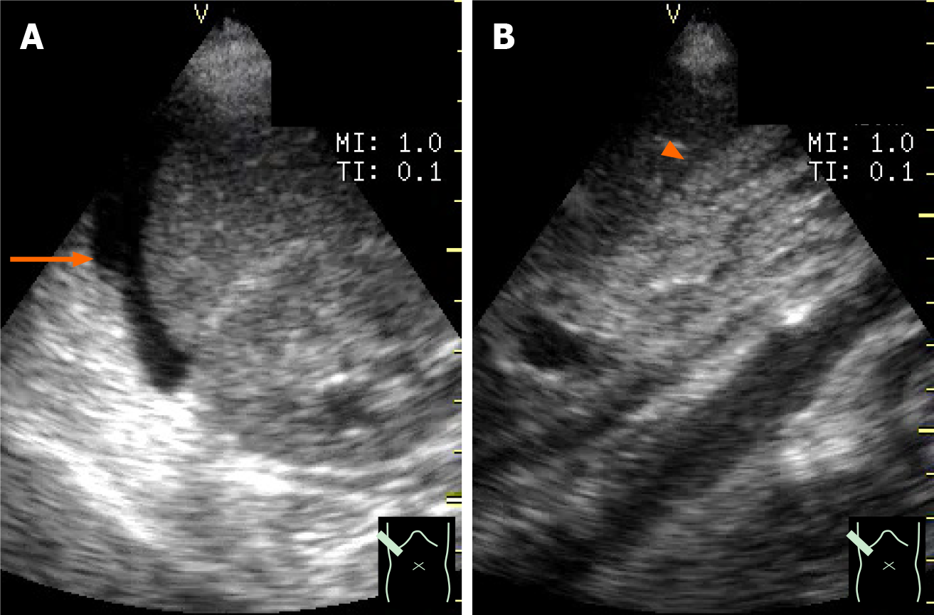
Figure 12 Case presentation 3 of asymptomatic patient with acute hepatitis.
The patient was asymptomatic upon admission. A: Pocket-sized ultrasound performed as part of a physical examination during medical rounds revealed a small amount of ascites (arrow) around the liver; B: The gallbladder was collapsed (arrowhead). She was diagnosed with severe acute hepatitis, and an energic treatment began immediately.
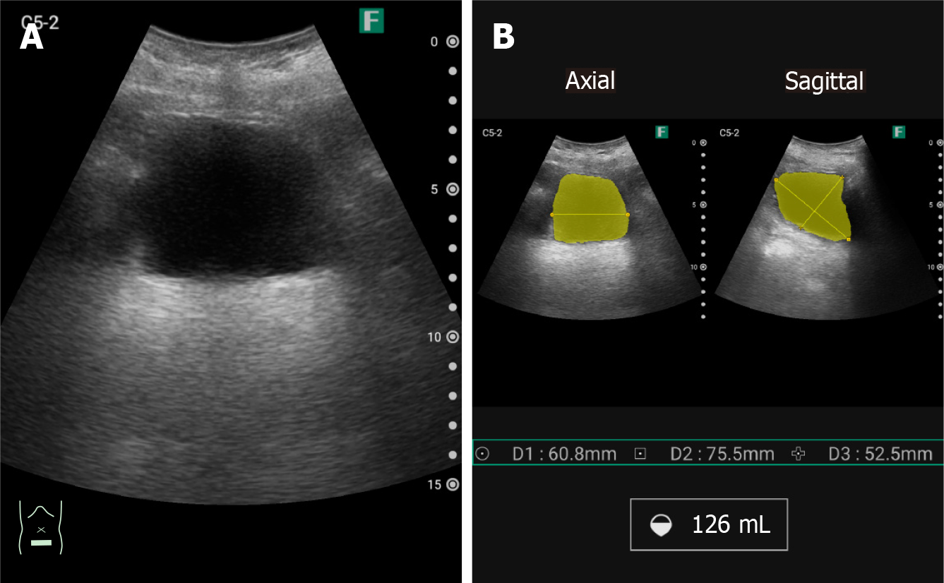
Figure 13 Built-in volume measurement application in pocket-sized ultrasound device.
This function is useful for evaluating urine volume in elderly patients. A: Scanning with pocket-sized ultrasound device; B: Urine volume display.
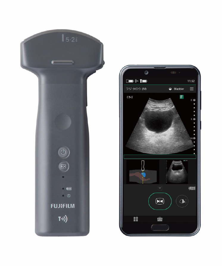
Figure 14 Built-in wireless function application in pocket-sized ultrasound device.
This function is particularly important when sending ultrasound information from distant locations (ambulance, etc.) or infection zones.
- Citation: Naganuma H, Ishida H. One-day seminar for residents for implementing abdominal pocket-sized ultrasound. World J Methodol 2021; 11(4): 208-221
- URL: https://www.wjgnet.com/2222-0682/full/v11/i4/208.htm
- DOI: https://dx.doi.org/10.5662/wjm.v11.i4.208


