Copyright
©The Author(s) 2021.
World J Methodol. Jul 20, 2021; 11(4): 160-186
Published online Jul 20, 2021. doi: 10.5662/wjm.v11.i4.160
Published online Jul 20, 2021. doi: 10.5662/wjm.v11.i4.160
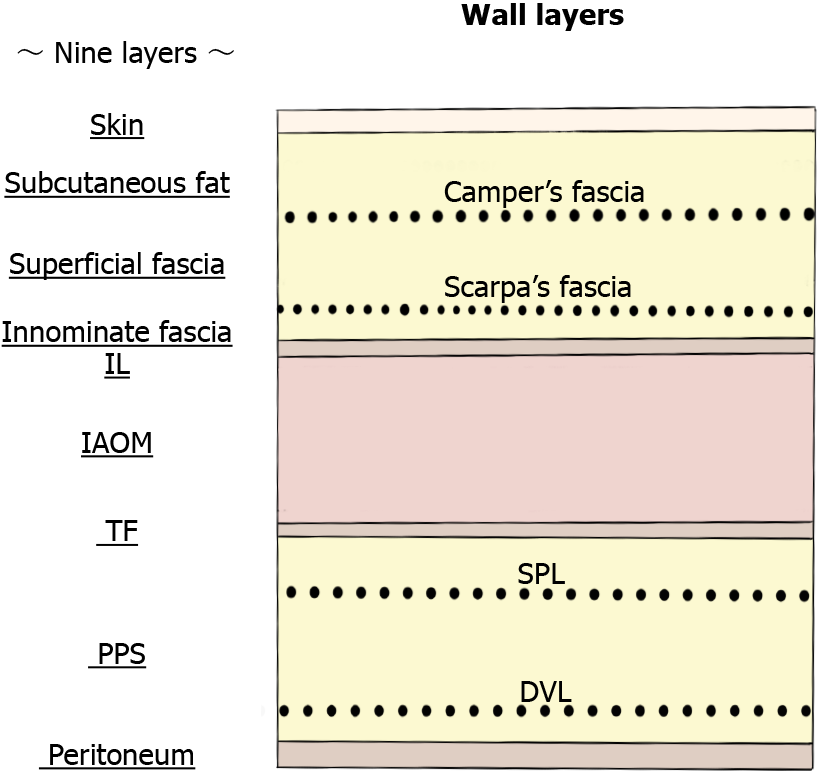
Figure 1 Wall layers.
The abdominal wall at the groin contains the following components: skin, subcutaneous fat, superficial fasciae (Camper’s and Scarpa’s fasciae), innominate (unnamed or untitled) fascia, IL, IAOM, TF, PPS [SPL (anterior subperitoneal fascia) and DVL (posterior subperitoneal fascia)], and peritoneum. DVL: Deeper visceral layer; IAOM: Internal abdominal oblique muscle; IL: Inguinal ligament; PPS: Preperitoneal space; SPL: Superficial parietal layer; TF: Transversalis fascia.
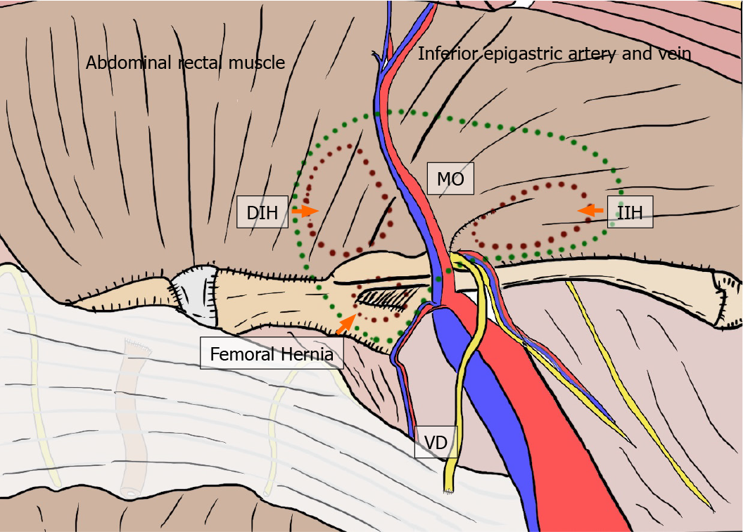
Figure 2 Myopectineal orifice.
The oval-shaped myopectineal orifice (green dotted circle) is the origin of all groin hernias (brown dotted circles). DIH: Direct inguinal hernia; IIH: Indirect inguinal hernia; MO: Myopectineal orifice; VD: Vas deferens.
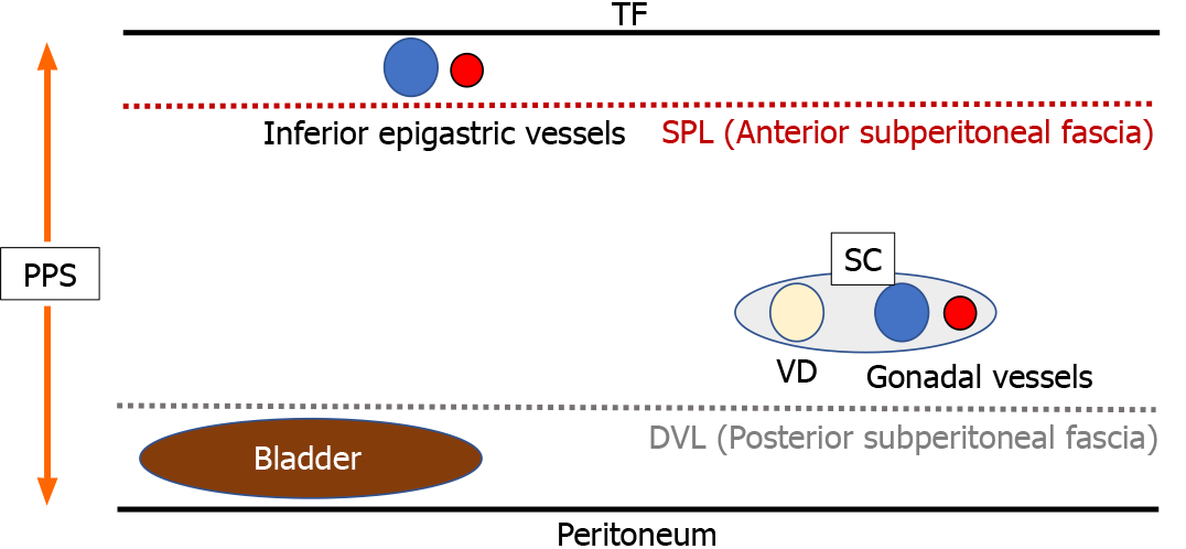
Figure 3 Preperitoneal space.
DVL: Deeper visceral layer; PPS: Preperitoneal space; SC: Spermatic cord; SPL: Superficial parietal layer; TF: Transversalis fascia; VD: Vas deferens.
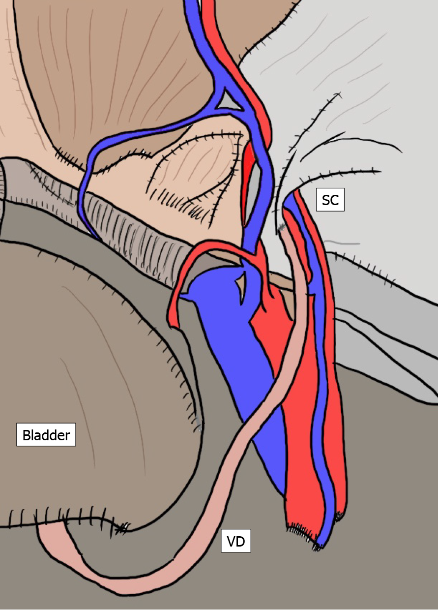
Figure 4 Vas deferens, spermatic cord and bladder.
The vas deferens courses as the “preperitoneal loop” in the deeper visceral layer (DVL). The bladder exists in the DVL. DVL: Deeper visceral layer; PPS: Preperitoneal space; SC: Spermatic cord; SPL: Superficial parietal layer; TF: Transversalis fascia; VD: Vas deferens.
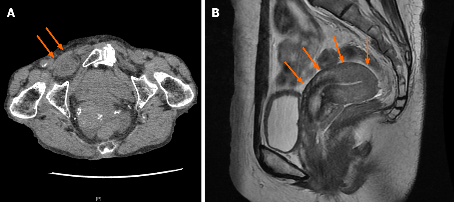
Figure 5 Actual image findings.
Actual image findings of obturator hernia in computed tomography (A, orange arrows) and retroflexion of the uterus in magnetic resonance imaging (B, orange arrows) are shown, respectively.
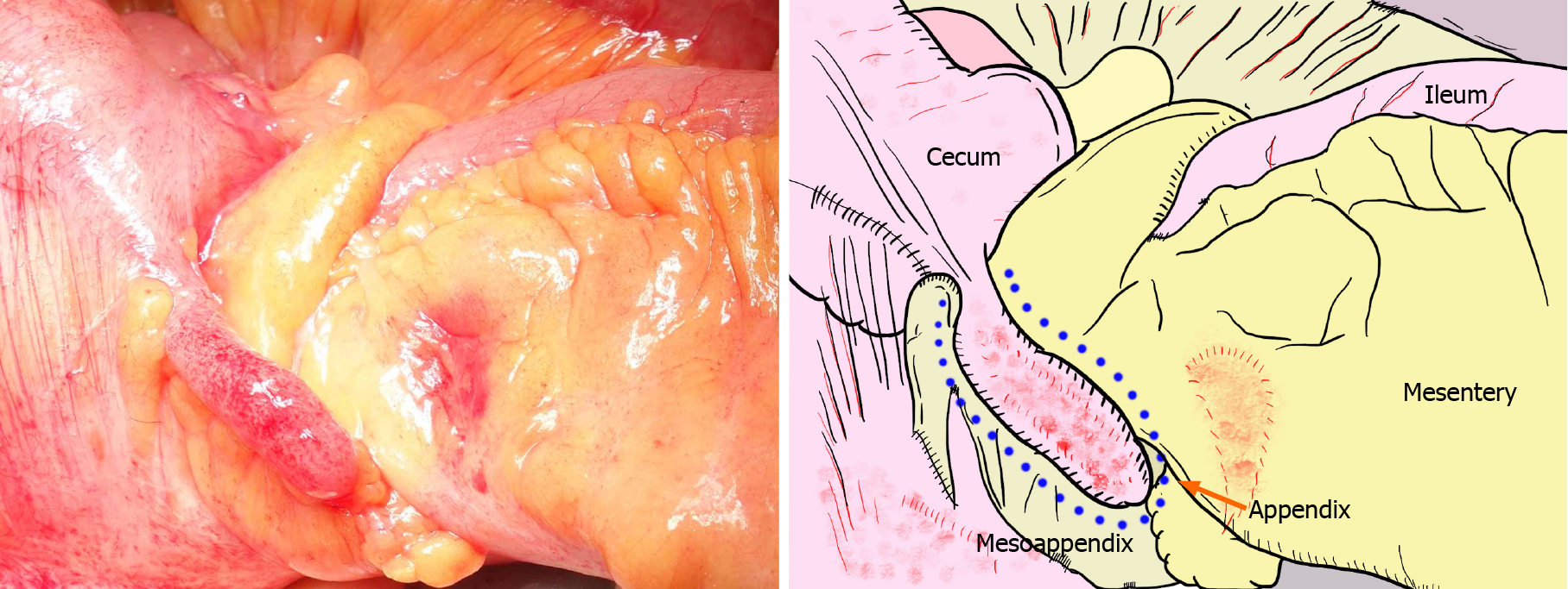
Figure 6 Amyand’s hernia.
Amyand’s hernia is considered as an inguinal hernia that traps the appendix. In patients who exhibit a giant hernia involving incarceration of the ileocecal portion of the intestinal tract, only the appendix does not recover from ischemic changes, despite resolution of the strangulation.
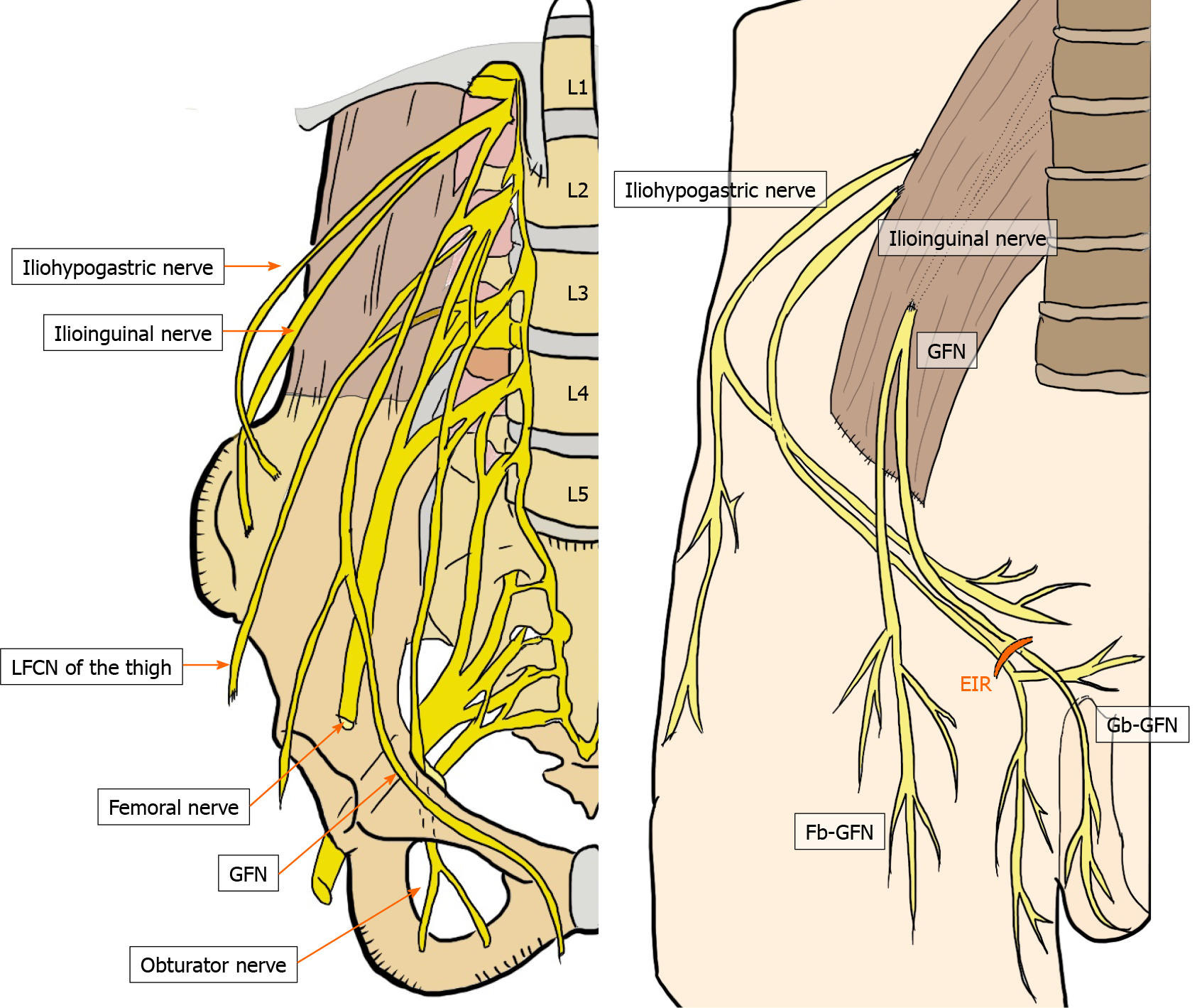
Figure 7 Topographic nerves located in the groin.
These six nerves of interest are the iliohypogastric, ilioinguinal, femoral (including the anterior cutaneous branch), genitofemoral (femoral and genital branches), lateral femoral cutaneous of the thigh, and obturator nerves. EIR: External inguinal ring; GFN: Genitofemoral nerve; Fb-GFN: The femoral branch of the GFN; Gb-GFN: The genital branch of the GFN; LFCN: Lateral femoral cutaneous nerve.
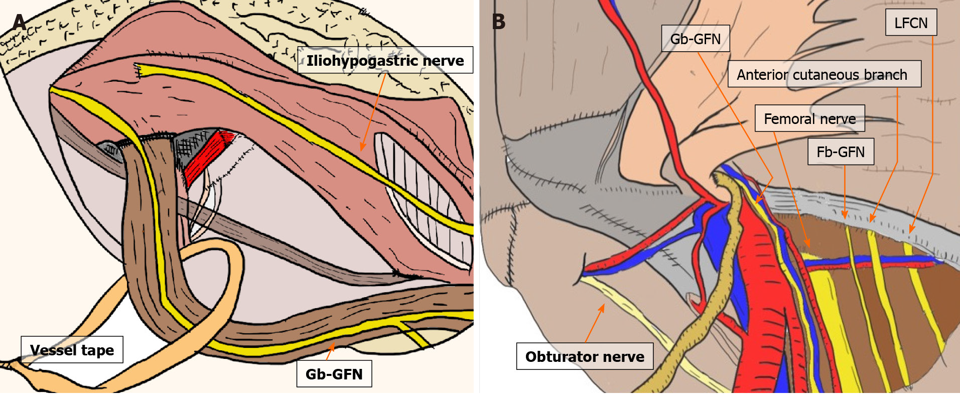
Figure 8 Topographic nerves located in the groin.
Respective anterior (A) and laparoscopic (B) views are shown. Fb-GFN: The femoral branch of the genitofemoral nerve; Gb-GFN: The genital branch of the genitofemoral nerve; LFCN: Lateral femoral cutaneous nerve.
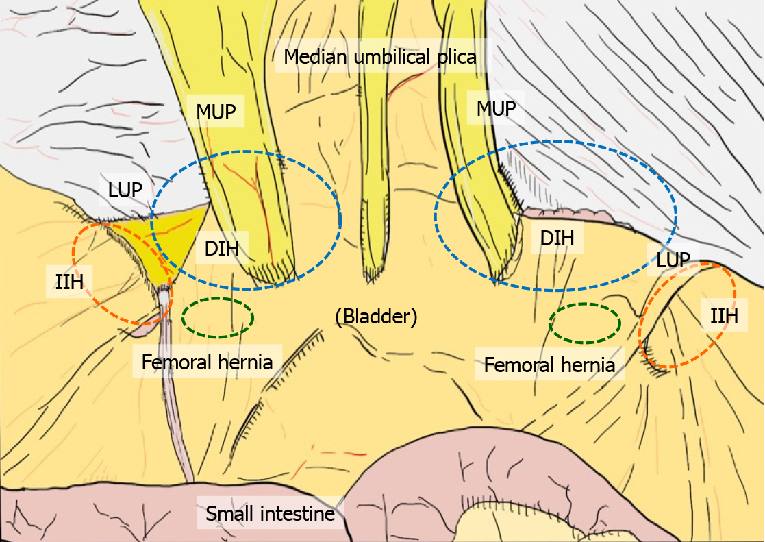
Figure 9 Plicae and fossae.
Plicae create three flat fossae recognizable on each side, corresponding to possible hernia defects. Hernia presentation can be more easily evaluated by a laparoscopic view than by an endoscopic or anterior view. DIH: Direct inguinal hernia; IIH: Indirect inguinal hernia; LUP: Lateral umbilical plica; MUP: Medial umbilical plica.
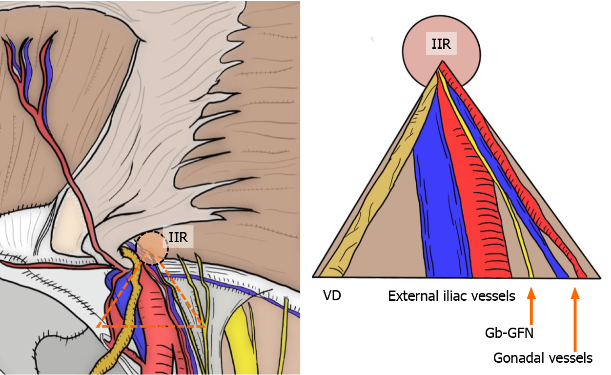
Figure 10 “Triangle of doom”.
The “triangle of doom” (orange dotted triangle) delineates the region between the VD and spermatic vessels. Currently, the “triangle of doom” is regarded as an inverted V-shaped area bound laterally by the gonadal vessels (in both sexes) and medially by the VD (in men and boys) or RL (in women and girls). Gb-GFN: The genital branch of the genitofemoral nerve; IIR: Internal inguinal ring; RL: Round ligament; VD: Vas deferens.
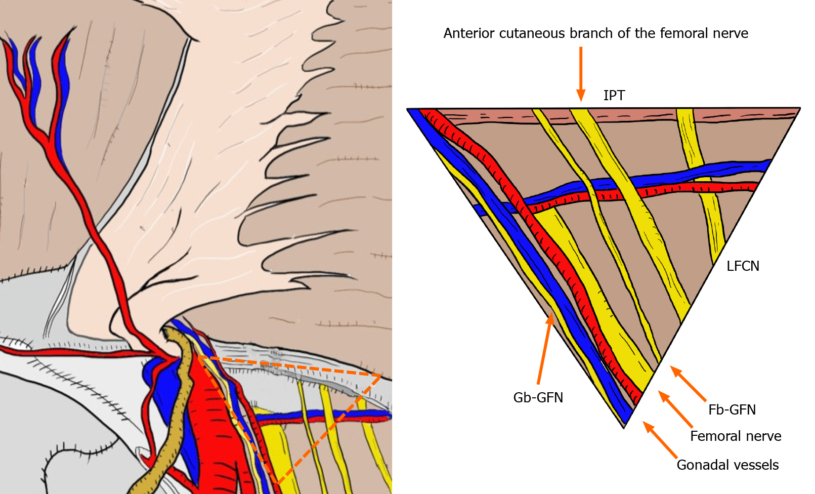
Figure 11 “Triangle of pain”.
The “triangle of pain” (orange dotted triangle) is defined as the area lateral to the testicular vessels and inferior to the iliopubic tract. The “triangle of pain” involves the femoral branch of the genitofemoral nerve, lateral femoral cutaneous nerve, femoral nerve, and anterior cutaneous branch of the femoral nerve. Subtle injury (or greater) to the nerves located within the “triangle of pain” is a risk factor for intractable pain. Fb-GFN: The femoral branch of the genitofemoral nerve; Gb-GFN: The genital branch of the genitofemoral nerve; IPT: Iliopubic tract; LFCN: Lateral femoral cutaneous nerve.
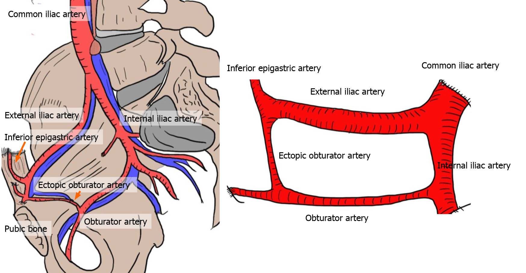
Figure 12 Corona mortis.
The corona mortis is classically defined as the arterial anastomosis between the obturator and the inferior epigastric arteries by means of the ectopic obturator artery. The existence of the obturator artery results in annular communication among the inferior epigastric, common iliac, internal iliac, external iliac, and obturator arteries. Brisk bleeding is difficult to control because of the dual vascular supply from the obturator and iliac vessels.
- Citation: Hori T, Yasukawa D. Fascinating history of groin hernias: Comprehensive recognition of anatomy, classic considerations for herniorrhaphy, and current controversies in hernioplasty. World J Methodol 2021; 11(4): 160-186
- URL: https://www.wjgnet.com/2222-0682/full/v11/i4/160.htm
- DOI: https://dx.doi.org/10.5662/wjm.v11.i4.160


