Copyright
©The Author(s) 2021.
World J Methodol. May 20, 2021; 11(3): 81-87
Published online May 20, 2021. doi: 10.5662/wjm.v11.i3.81
Published online May 20, 2021. doi: 10.5662/wjm.v11.i3.81
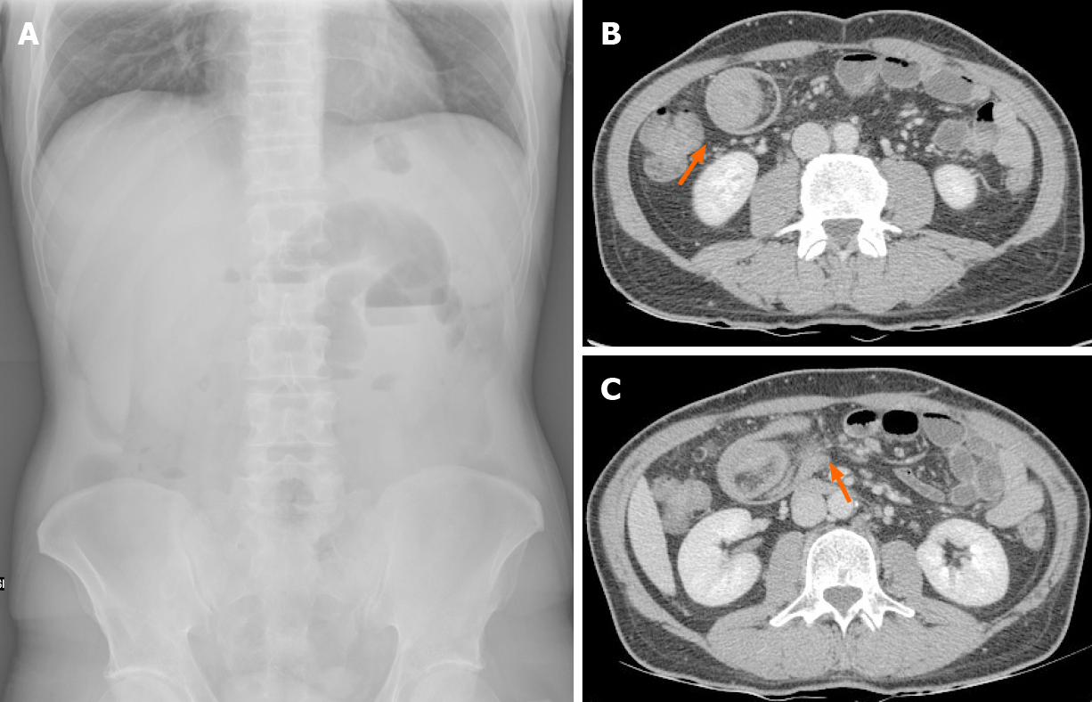
Figure 1 Inflammatory fibroid polyp of the small intestine.
A 43-year-old male presenting with abdominal pain and vomiting. A: Abdomen X-ray showed signs of intestinal obstruction with hydro-air levels in the upper quadrants; B: Computed tomography scan confirmed bowel obstruction with presence of “target sign” (orange arrow); C: Mesenteric fat and blood vessels are visible (orange arrow). Surgical resection revealed an inflammatory fibroid polyp of the ileum.
Figure 2 Ileocecal valve adenocarcinoma.
A 56-year-old female presenting with right iliac fossa pain. A: Ultrasound scan revealed “target sign”; B and C: Computed tomography scan confirmed ielo-colic intussusception, with no signs of bowel obstruction [orange arrow, horizontal (B) and coronal (C)]. Surgical resection revealed an ileocecal valve adenocarcinoma (pT2 N0).
- Citation: Panzera F, Di Venere B, Rizzi M, Biscaglia A, Praticò CA, Nasti G, Mardighian A, Nunes TF, Inchingolo R. Bowel intussusception in adult: Prevalence, diagnostic tools and therapy. World J Methodol 2021; 11(3): 81-87
- URL: https://www.wjgnet.com/2222-0682/full/v11/i3/81.htm
- DOI: https://dx.doi.org/10.5662/wjm.v11.i3.81


