Editorial Open Access
Copyright ©The Author(s) 2021. Published by Baishideng Publishing Group Inc. All rights reserved.
World J Transl Med. Mar 15, 2021; 9(1): 1-10
Published online Mar 15, 2021. doi: 10.5528/wjtm.v9.i1.1
Machine intelligence for precision oncology
Nelson S Yee, Department of Medicine, The Pennsylvania State University College of Medicine, Penn State Cancer Institute, Penn State Health Milton S. Hershey Medical Center, Hershey, PA 17033-0850, United States
ORCID number: Nelson S Yee (0000-0002-1457-9047).
Author contributions: Yee NS performed research and wrote the paper.
Conflict-of-interest statement: Dr. Yee reports grants from Ipsen Biopharmaceuticals, other from Caris Life Sciences, other from Novartis, outside the submitted work.
Open-Access: This article is an open-access article that was selected by an in-house editor and fully peer-reviewed by external reviewers. It is distributed in accordance with the Creative Commons Attribution NonCommercial (CC BY-NC 4.0) license, which permits others to distribute, remix, adapt, build upon this work non-commercially, and license their derivative works on different terms, provided the original work is properly cited and the use is non-commercial. See: http://creativecommons.org/Licenses/by-nc/4.0/
Corresponding author: Nelson S Yee, BPharm, FACP, MD, PhD, Associate Professor, Attending Doctor, Department of Medicine, The Pennsylvania State University College of Medicine, Penn State Cancer Institute, Penn State Health Milton S. Hershey Medical Center, 500 University Drive, Hershey, PA 17033-0850, United States. nyee@pennstatehealth.psu.edu
Received: October 19, 2020
Peer-review started: October 19, 2020
First decision: November 16, 2020
Revised: December 22, 2020
Accepted: March 1, 2021
Article in press: March 1, 2021
Published online: March 15, 2021
Processing time: 139 Days and 16.8 Hours

Abstract

Despite various advances in cancer research, the incidence and mortality rates of malignant diseases have remained high. Accurate risk assessment, prevention, detection, and treatment of cancer tailored to the individual are major challenges in clinical oncology. Artificial intelligence (AI), a field of applied computer science, has shown promising potential of accelerating evolution of healthcare towards precision oncology. This article focuses on highlights of the application of data-driven machine learning (ML) and deep learning (DL) in translational research for cancer diagnosis, prognosis, treatment, and clinical outcomes. ML-based algorithms in radiological and histological images have been demonstrated to improve detection and diagnosis of cancer. DL-based prediction models in molecular or multi-omics datasets of cancer for biomarkers and targets enable drug discovery and treatment. ML approaches combining radiomics with genomics and other omics data enhance the power of AI in improving diagnosis, prognostication, and treatment of cancer. Ethical and regulatory issues involving patient confidentiality and data security impose certain limitations on practical implementation of ML in clinical oncology. However, the ultimate goal of application of AI in cancer research is to develop and implement multi-modal machine intelligence for improving clinical decision on individualized management of patients.

Key Words: Artificial intelligence; Deep learning; Machine learning; Precision oncology; Radiomics; Radiogenomics

Core Tip: Artificial intelligence represents the future of healthcare particularly precision oncology for prevention, detection, risk assessment, and treatment of cancer. Application of machine learning- and deep learning-based algorithms in translational research has been demonstrated to improve accuracy of cancer diagnosis and anti-cancer drug development. Multi-disciplinary collaboration with resolution of ethical and regulatory issues of multi-modal machine intelligence are indicated for implementation of computer-assisted clinical decision on individualized patient management.


INTRODUCTION

Precision medicine is the new frontier of healthcare and medical research, and it has been mainly implemented in oncology. Precision oncology can be defined as an approach for treatment and prevention of cancer with the individual variability in genes, environment, and behavior taken into account. Accurate risk assessment, prevention, detection, and treatment of cancer tailored to the individual are major challenges in clinical oncology. Despite various advances in basic, translational, and clinical cancer research, the incidence and mortality rates of malignant diseases have remained high[1,2]. Artificial intelligence (AI), a field of applied computer science, has shown promising potential of accelerating towards the goal of precision oncology. Application of AI in oncology involves integrative analysis of “big cancer data” such as digitized images, multi-omics, clinical datasets, and population health.

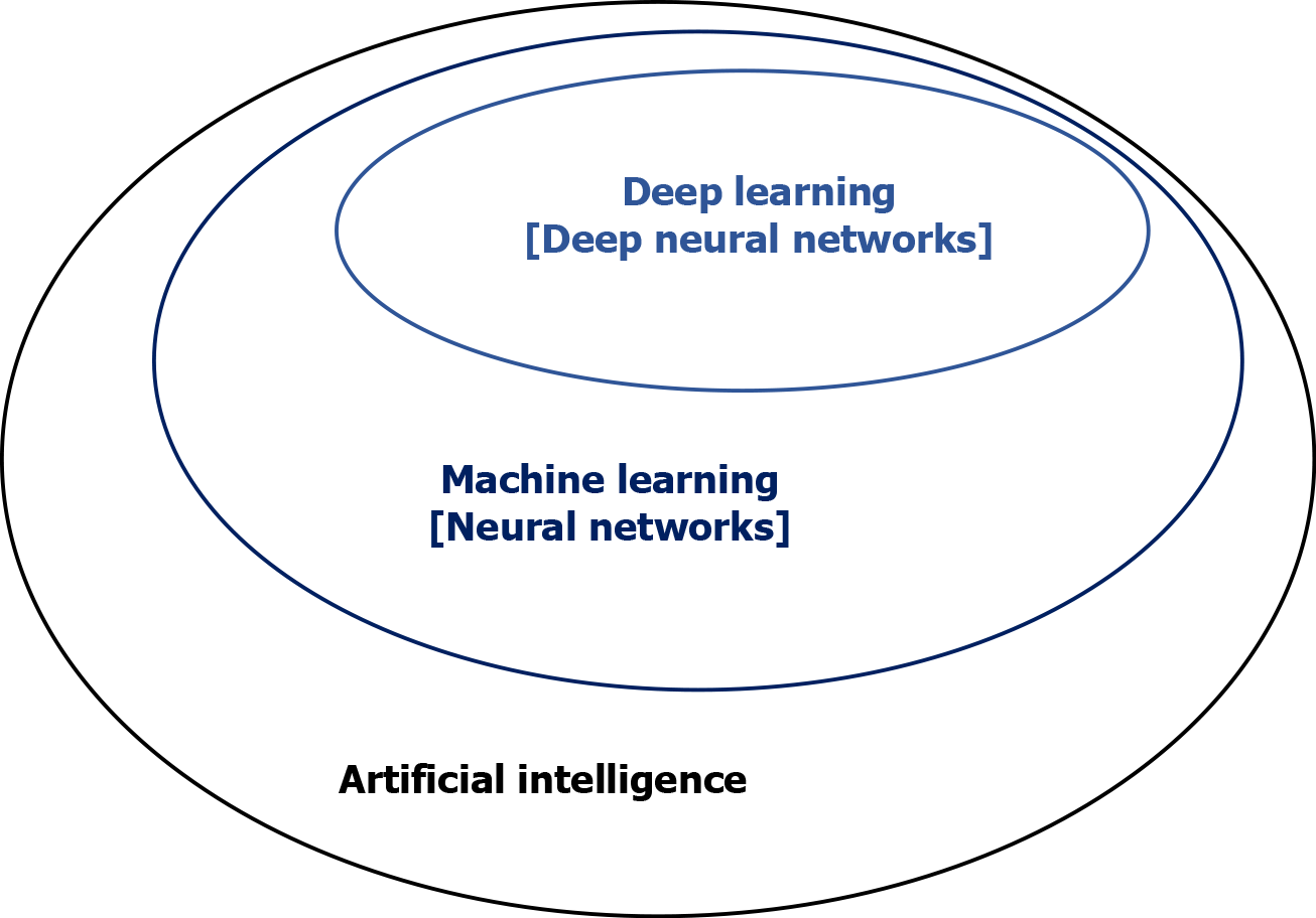
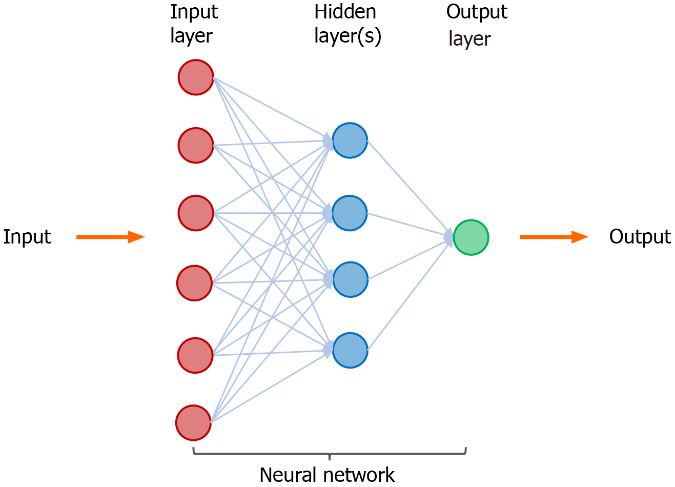
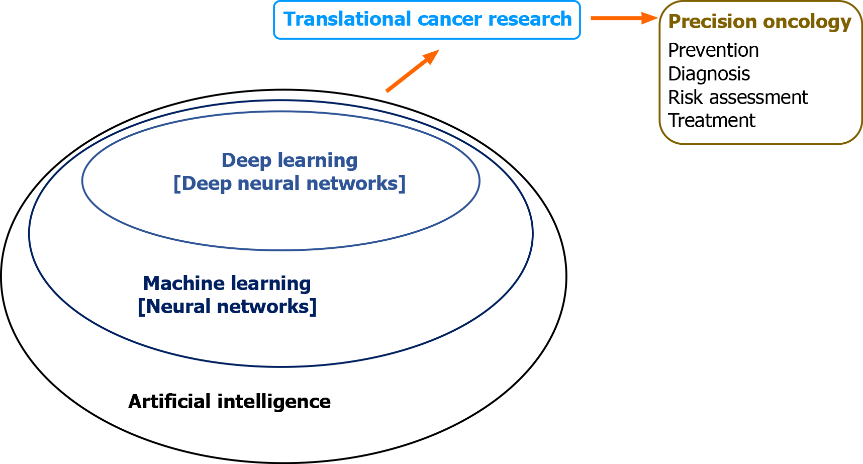
With the advent of electronic health records, bio-banking, multi-omics, and digitized radiographic and histological images, we have entered the era of big data and team science. AI has emerged as a powerful technology that will transform healthcare by multi-disciplinary research to capture and analyze large pools of data[3,4]. Application of AI in translational cancer research has shown its potential for advancing diagnosis, prognostication, and treatment[5]. This is accomplished by integration and analysis of large data sets and generating algorithms-based prediction models. Machine learning (ML) is a branch of AI that applies statistical methods to detect patterns within datasets[6]. Deep learning (DL), characterized by deep artificial neural network, is a sub-branch of ML that utilizes the capability of multi-layered networks[7] (Figures 1 and 2). Application of ML and DL approaches has been demonstrated to advance translational cancer research in various aspects. These include detection and classification of tumor subtypes, diagnosis of cancer, assessment of cancer risk, prediction of clinical outcomes, discovery of cancer biomarkers, repurposing of drugs for cancer treatment, and predicting drug response of tumors.

Figure 1
Figure 1 Artificial intelligence, machine learning, and deep learning. Artificial intelligence is a field of applied computer science that mimics human cognition to complete a task. Machine learning is a branch of artificial intelligence that manually extract features from input data to create a model that categorizes the object. Neural network is a set of machine-based learning algorithms to learn labeled datasets and perform classification tasks, and it comprises an input layer, a hidden layer of interconnected nodes, and an output layer. Deep learning is a machine learning technique that uses neural network architectures, and the term “deep” refers to the number of hidden layers (more than three) in the neural network.
Figure 2
Figure 2 A neural network in machine learning. Neural network is a set of machine-based learning algorithms, and it comprises an input layer (red circles), a hidden layer of interconnected nodes (blue circles), and output layer (green circle). The function of a neural network is to extract and process features from labeled datasets and perform classification tasks. A representative hidden layer of interconnected nodes (blue circles) is shown. A deep learning node (blue circle) is a computational unit that combines input data with weights (assigned significance) and generate an output layer. For deep learning, a neural network contains more than three hidden layers.
ML IN RADIOLOGICAL IMAGES OF CANCER

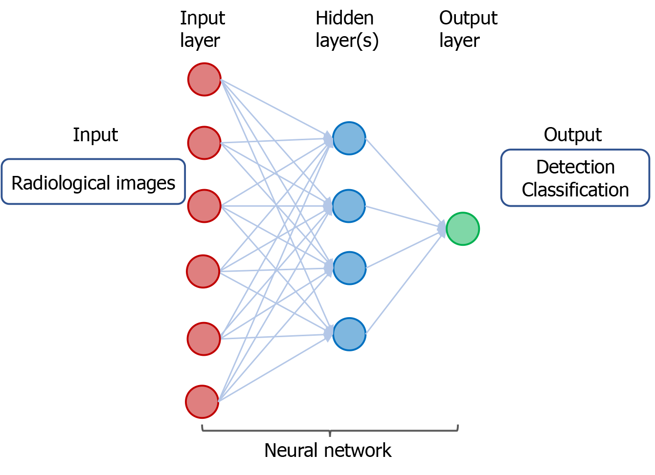
Classification and early detection of cancer are crucial for accurate diagnosis and treatment with curative intent. In “radiomics”, the data based on algorithm for extracting and analyzing features from medical images enable improvement of accuracy of cancer diagnosis, prognostication, and clinical prediction[8]. Advances have been made in research of DL with convolutional neural networks (CNN), an algorithm to process and differentiate images, in cancer imaging and help facilitate accurate classification and detection of cancer[9] (Figure 3).

Figure 3
Figure 3 A neural network in deep learning of radiological images. The input consists of data derived from radiological images from individuals with cancer or without cancer. The output includes detection and classification of tumors.

DL with CNN and its variants has been applied for classification and detection of cancer in different organs. Several studies using DL in radiological images as input data are described as follows. Using a dataset of about 130000 clinical images of skin lesions, a trained CNN is capable of a dermatologist-level classification of keratinocyte carcinoma and malignant melanoma[10]. As shown in a systematic review of eleven studies, CNN enables accurate diagnosis of hepatocellular carcinoma by recognizing specific features in computed tomographic (CT) or magnetic resonance images[11]. Based on retrospective datasets of 2652 digital mammography, 653 of which showed malignancy, the AI system using DL CNN algorithms to detect calcifications and soft tissue lesions showed an accuracy for detection of breast cancer comparable to an average breast radiologist[12]. Using two independent datasets for training and validating the AI algorithm, the accuracy of a DL-based model for screening breast cancer by mammography is superior to that of expert radiologists, with the area under the receiver operating characteristic curve (AUC-ROC) for the AI system greater than that for the average radiologist by 11.5%[13]. By combining 3-dimensional deep CNN with cloud computing for analysis of the datasets of lung nodules on chest CT imaging, lung cancer can be accurately detected with a sensitivity of 98.7% at 1.97 false positives per scan[14].

Results of these studies demonstrate the power of AI using DL CNN algorithms for accurately detecting cancer in different organs (Figure 3). Future investigation by clinical trials is indicated to improve the accuracy and efficiency of cancer detection using the AI systems. Multi-disciplinary and coordinated research efforts are necessary to determine how the DL-based models can be potentially integrated into clinical practice.

ML IN HISTOLOGICAL IMAGES OF CANCER

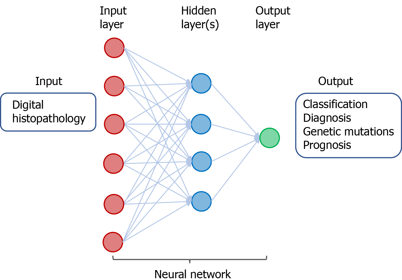
Microscopic analysis of tumor histopathology with immunohistochemistry has been the standard practice for diagnosis and grading of cancer. With the advent of scanning technology, digitization of whole slide images of biopsied or resected tumor specimens has enabled computer-assisted analysis to improve accuracy and efficiency of diagnosis. ML/DL for analysis of digitalized images of tumor histopathology has been demonstrated to have potential for improving diagnosis of cancer, identifying tumor and lymph node metastasis, predicting genetic mutation and clinical outcomes[15] (Figure 4).

Figure 4
Figure 4 A neural network in deep learning of digital histopathology. The input consists of digital histopathological data derived from individuals with or without cancer. The output includes classification and diagnosis of cancer as well as predicting genetic mutations and prognosis of patients with cancer.

Here are a few examples that illustrate the power of ML in digitized images of cancer for improving accuracy of pathological diagnosis. Using DL with CNN technique for analysis of biopsied tissue specimens, prostate cancer as well as micro- and macro-metastases of breast cancer in sentinel lymph nodes could be automatically identified without the need for immunohistochemistry[16]. Similarly, using DL-based approaches to train a CNN to discriminate tumor from normal tissue, metastatic breast cancer could be automatically detected in images of biopsied sentinel lymph nodes[17,18].

Moreover, DL-based algorithms can be trained to analyze histopathological images and predict mutation and clinical outcomes. A deep CNN was trained to analyze whole slide images obtained from The Cancer Genome Atlas (https://portal.gdc.cancer.gov). The DL method could automatically classify the tissues as lung adenocarcinoma, squamous cell carcinoma, or normal lung tissue. In addition, the trained CNN could accurately predict some of the commonly mutated genes in lung adenocarcinoma including EGFR, KRAS, TP53, FAT1, SETBP1, and STK11[19]. A novel DL-based approach to train a deep network was used to analyze digitized tissue microarray specimens of colorectal cancer from 420 patients, along with their clinicopathological features and clinical outcomes. Results of this study show that DL-based prediction of 5-year disease specific survival is superior to that by visual evaluation of histology by expert pathologists[20].

DL with CNN approaches have shown promising potential for improving digitized histopathology-based diagnostics (Figure 4). In order to apply for diagnosis and classification of cancer in clinical practice, the utility of ML/DL in analyzing digital tumor histopathology will need to be assessed and validated in prospective clinical trials involving a large number of patients. Furthermore, combination of ML in digitized histopathology with other datasets such as tumor omics can enhance the predictive capability and accuracy.

ML IN MULTI-OMICS DATASETS OF CANCER

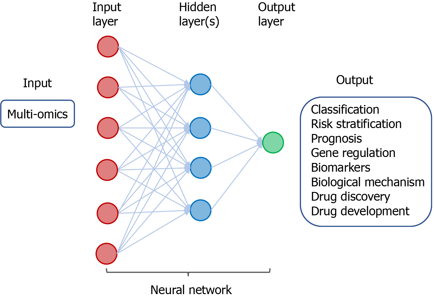
Research on molecular characterization of tumors has generated a wealth of data on genetic and epigenetic alterations that control carcinogenesis[21]. Tissue-based omics have yielded tremendous number of clinically useful cancer biomarkers and targets. These include data derived from genomics, epigenomics, transcriptomics, proteomics, metabolomics, phenomics, and metagenomics [Genomic Data Commons Data Portal (https://portal.gdc.cancer.gov)][22]. These omics data have been used to classify tumor types, identify and develop cancer biomarkers, and drug discovery and development. Moreover, ML/DL may help improve the efficiency and accuracy omics-based therapeutic strategies[23](Figure 5).

Figure 5
Figure 5 A neural network in deep learning of cancer-derived multi-omics. The input consists of multi-omics data derived from various cancers. The output includes classification and risk stratification of cancer, predicting prognosis, and investigating gene regulation/biomarkers/biological mechanism, as well as drug discovery and development.

DL algorithms for analysis of omics data have been demonstrated to facilitate classification and detection of cancer as well as stratification of risk in patients with cancer. Using a DL approach, termed Stacked Denoising Autoencoder, to extract features from RNA sequencing (RNA-seq) expression in The Cancer Genome Atlas (TCGA) database, breast tissues can be classified into cancer or non-cancer and the involved genes can be identified as potential cancer biomarkers[24]. By application of CNN for analysis of RNA-seq data in Pan-Cancer Atlas, tissue samples have been classified with accuracy into 33 different types of cancer[25]. Besides gene expression data, DL analysis of epigenetics data particularly DNA methylation in the context of CpG islands has also been shown to classify cancer types. A deep neural network (DNN) was developed to extract the deep features of DNA methylation data, and this method can differentiate patients with breast cancer from healthy individuals[26]. Similarly, a CNN-based DL model can classify different types of cancer by analysis of the patterns of DNA methylation[27]. Furthermore, an advanced DNN-based model, DeepGene, was developed to analyze somatic point mutation data, and it was demonstrated to improve classification of 12 selected types of cancer[28]. Recently, the power of DL and traditional ML methods in cancer classification using TCGA datasets was compared, and results of the study indicate that the DL method, termed Multi-Layer Perceptions, outperforms the other approaches in discrimination of samples with cancer from non-cancer[29].

In addition to classification of cancer types, DL and ML have been exploited to predict patient prognosis and investigate gene regulation. By DL-based analysis of multi-omics data, including RNA-seq, microRNA sequencing (miRNA-seq), and DNA methylation data, patients with hepatocellular carcinoma can be classified into subgroups with difference in survival[30]. DL autoencoder algorithm for analysis of multi-omics datasets comprising mRNA, miRNA, and DNA methylation from TCGA can also predict the survival subtypes of patients with urinary bladder cancer[31]. Besides, ML-based integrative analysis of multi-omics data on the cloud has been demonstrated to improve the accessibility and productivity of cancer research for discovery of gene regulatory subnetwork, analysis of disease subtype, analysis of survival, prediction of clinical outcome, and visualization of multi-omics results[32].

By application of DL for integrative analysis of omics data, prediction models can be generated for discovering biomarkers and repurposing drugs[33,34]. In a proof-of-principle study using transcriptomic profiles of cancer cells treated with a variety of drugs at different concentrations, DNNs were trained to classify those drugs into therapeutic categories[35]. Using chemical and genetic information, a DL approach was shown to model and predict synergistic actions of novel combinations of anti-cancer drugs[36]. A DNN-based framework, termed PADME (Protein And Drug Molecule interaction prediction), was developed to predict drug-target interaction with input of information on compounds and protein[37].

Application of ML/DL in analyzing multi-omics has shown utility for classification of cancer and its risk stratification prognosis, demonstrated the power of investigating gene regulation, biomarkers, and biological mechanism, and created ample opportunity for drug discovery and development (Figure 5). Emerging studies have explored the potential of DL in omics-based training for prediction of tumor response to therapy, monitoring tumor response during treatment, and patient prognosis. Various bioinformatics tools have been developed and applied in the analysis of omics and inter-omics data in cancer. These approaches may improve diagnosis of breast cancer[38], enhance classification of cancer[39], identify cancer-associated sub-pathways[40], and provide insight into the oncogenic mechanisms and molecular biomarkers in malignant gliomas[41]. Further application of ML/DL in omics datasets in combination with other input data such as radiological images and digitized histopathology has been shown to enhance the power of AI for precision oncology.

MULTI-MODAL ML FOR PRECISION ONCOLOGY

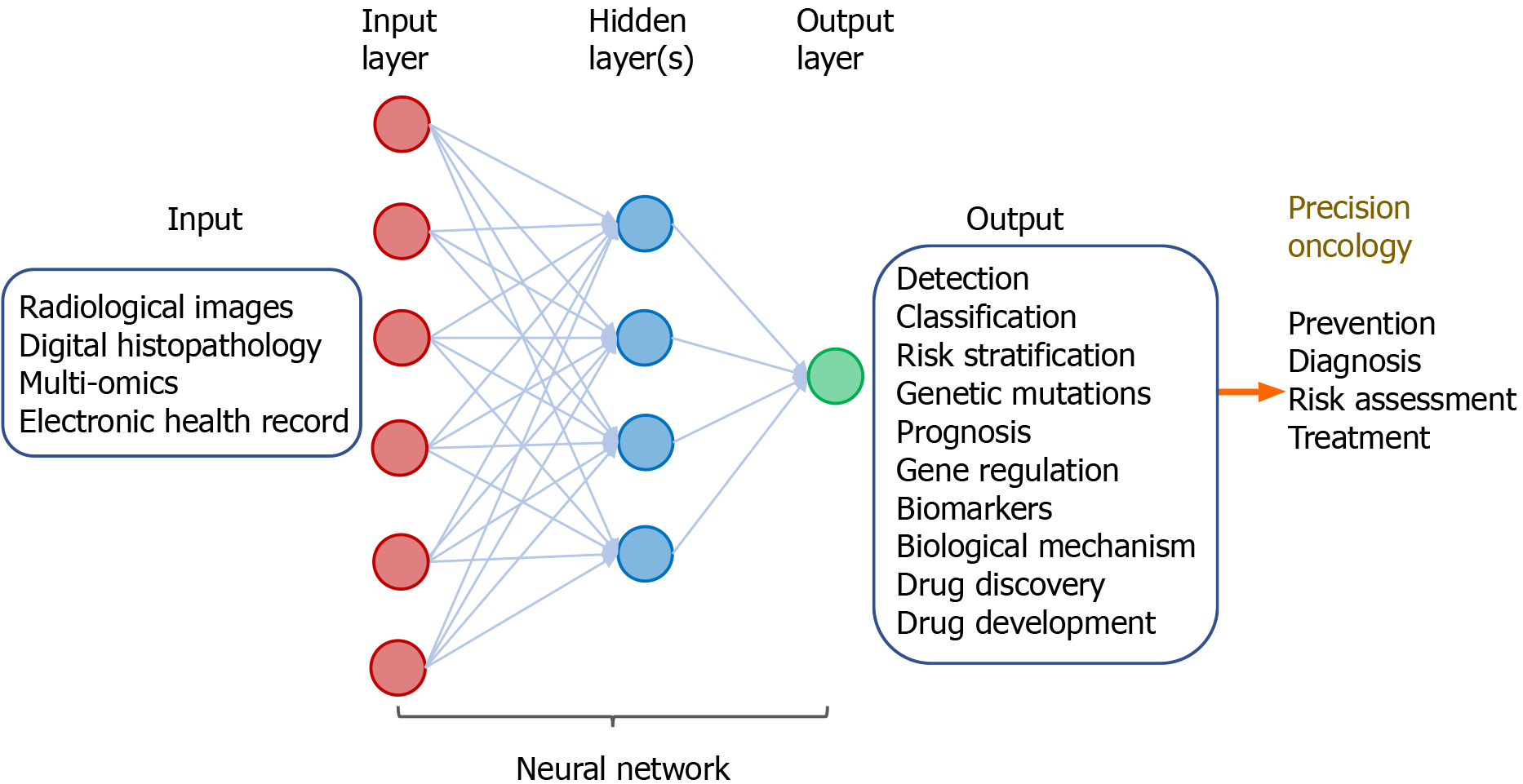
ML by integrative analysis of large data pools combining different types of inputs has been demonstrated to improve accuracy for prediction of diagnosis and clinical outcomes. This involves multi-modal ML in combination of radiological images, digitized histopathology, and omics in conjunction with electronic clinical data. These algorithm-based network models help support and facilitate clinical decision on diagnosis, prognostication, treatment, and patient stratification (Figure 6).

Figure 6
Figure 6 Multi-modal deep learning for precision oncology. The input comprises clinicopathological datasets from healthy individuals and patients with cancers. The output includes a variety of predicted outcomes that form the basis of precision oncology.

The power of multi-modal ML for precision oncology has been demonstrated by a “holomics” approach that combines medical images, histology, multi-omics, and clinical parameters[42]. By associating radiographic features with patterns of gene expression, a method known as “radiogenomics”, the algorithm-based data produce information on the underlying disease processes and enable prediction of molecular subtypes. Radiomic features can also been linked with other omics data such as proteomics, metabolomics, and immunomics. The radiomic biomarkers being generated can serve as surrogates that help facilitate diagnosis, prognostication, and prediction of tumor response to treatment[43]. In a recent article, a large number of studies in radiogenomics across a variety of tumor sites was reviewed[44]. These include tumors in the brain, lung, breast, ovary, liver, colon/rectum, prostate, and kidney. In these radiogenomics studies, the imaging features of the tumors can be linked with specific cancer genetic mutations. Results of these studies suggest imaging signatures can be developed as predictive biomarkers of genetic alterations in the tumor.

Besides radiogenomics, multi-modal ML of other types of input data have been combined to generate prediction models of clinical outcomes. A computational approach using DL-based CNN combines learning of digitized images of histopathology and genomic biomarkers along with clinical data of patients with glioma. This inter-modal ML-based algorithm has led to development of a predictive model for determining patient survival[45]. By combined analysis of radiomic features of prostate gland with radiologist’s evaluation, prostate specific antigen density, and digital rectal examination, models were developed to characterize prostatic lesions as benign, clinically significant or insignificant cancer. The ML-based models help facilitate selection of patients for MRI-guided biopsy for detection of prostate tumor[46].

Multi-modal ML approaches in combining radiomics and genomics as well as other omics have held great promise for improving capability and accuracy of prediction models in clinical oncology (Figure 6). Application of multi-modal ML for various aspects of translational cancer research is expected to continue to expand. The technical and personnel limitations of this evolving field will need to tackled and resolved. Standardization of ML-based tools along with concerted efforts through collaboration among clinicians and information technologists will help accelerate implementation of multi-modal ML in diagnosis and treatment of cancer.

CONCLUSION

Medical application of AI technology has been revolutionizing healthcare. ML and DL algorithms create powerful tools and opportunities for advancing translational cancer research. Accumulating evidence has begun to demonstrate the value for improving various aspects of clinical oncology such as diagnosis and treatment of cancer. In particular, advances have been made by DNN-based analysis of “big cancer data” towards the goal of precision oncology (Figure 7).

Figure 7
Figure 7 Machine intelligence in translational cancer research for precision oncology.

A number of hurdles will need to be resolved in order to move toward implementation of multi-modal ML in clinical practice. Limitations in radiomics may include inter-observer variability of data processing, reproducibility of radiomic features, tumor heterogeneity, and difference in radiomics approach among researchers[8,44]. Large infrastructural networks and platforms for collection, processing, storage, sharing, and accessing medical images, histopathology, and clinical data across institutions may impose challenges[47]. Due to access of personal information and cloud-based storage of data, ethical and regulatory issues concerning patient confidentiality and data security are non-trivial[48-50].

However, multi-modal ML approaches that integrate large datasets, including medical images, digitalized pathology, holomics, and clinical features will continue to evolve. Emerging applications of AI in oncology involve ML in selection of treatment[23], palliative care and hospice[51], and design of clinical trials[52]. Multi-disciplinary collaboration for development and adoption of multi-modal ML is expected to accelerate healthcare evolution towards precision oncology through computer-aided clinical decision on individualized management of patients.

Footnotes

Manuscript source: Invited manuscript

Specialty type: Medicine, research and experimental

Country/Territory of origin: United States

Peer-review report’s scientific quality classification

Grade A (Excellent): 0

Grade B (Very good): B

Grade C (Good): C, C

Grade D (Fair): 0

Grade E (Poor): 0

P-Reviewer: Tran B, Tsui SKW S-Editor: Gao CC L-Editor: A P-Editor: Wang LL

References
1.  Bray F, Ferlay J, Soerjomataram I, Siegel RL, Torre LA, Jemal A. Global cancer statistics 2018: GLOBOCAN estimates of incidence and mortality worldwide for 36 cancers in 185 countries. CA Cancer J Clin. 2018;68:394-424.  [PubMed]  [DOI]  [Cited in This Article: ]  [Cited by in Crossref: 53206]  [Cited by in F6Publishing: 53776]  [Article Influence: 7682.3]  [Reference Citation Analysis (125)]
2.  Siegel RL, Miller KD, Jemal A. Cancer statistics, 2020. CA Cancer J Clin. 2020;70:7-30.  [PubMed]  [DOI]  [Cited in This Article: ]  [Cited by in Crossref: 12667]  [Cited by in F6Publishing: 14679]  [Article Influence: 2935.8]  [Reference Citation Analysis (4)]
3.  Yu KH, Beam AL, Kohane IS. Artificial intelligence in healthcare. Nat Biomed Eng. 2018;2:719-731.  [PubMed]  [DOI]  [Cited in This Article: ]  [Cited by in Crossref: 732]  [Cited by in F6Publishing: 945]  [Article Influence: 135.0]  [Reference Citation Analysis (0)]
4.  Matheny ME, Whicher D, Thadaney Israni S. Artificial Intelligence in Health Care: A Report From the National Academy of Medicine. JAMA. 2020;323:509-510.  [PubMed]  [DOI]  [Cited in This Article: ]  [Cited by in Crossref: 175]  [Cited by in F6Publishing: 209]  [Article Influence: 41.8]  [Reference Citation Analysis (0)]
5.  Huang S, Yang J, Fong S, Zhao Q. Artificial intelligence in cancer diagnosis and prognosis: Opportunities and challenges. Cancer Lett. 2020;471:61-71.  [PubMed]  [DOI]  [Cited in This Article: ]  [Cited by in Crossref: 133]  [Cited by in F6Publishing: 246]  [Article Influence: 41.0]  [Reference Citation Analysis (1)]
6.  Rajkomar A, Dean J, Kohane I. Machine Learning in Medicine. N Engl J Med. 2019;380:1347-1358.  [PubMed]  [DOI]  [Cited in This Article: ]  [Cited by in Crossref: 1274]  [Cited by in F6Publishing: 1416]  [Article Influence: 236.0]  [Reference Citation Analysis (0)]
7.  LeCun Y, Bengio Y, Hinton G. Deep learning. Nature. 2015;521:436-444.  [PubMed]  [DOI]  [Cited in This Article: ]  [Cited by in Crossref: 36149]  [Cited by in F6Publishing: 18679]  [Article Influence: 1867.9]  [Reference Citation Analysis (0)]
8.  Lambin P, Leijenaar RTH, Deist TM, Peerlings J, de Jong EEC, van Timmeren J, Sanduleanu S, Larue RTHM, Even AJG, Jochems A, van Wijk Y, Woodruff H, van Soest J, Lustberg T, Roelofs E, van Elmpt W, Dekker A, Mottaghy FM, Wildberger JE, Walsh S. Radiomics: the bridge between medical imaging and personalized medicine. Nat Rev Clin Oncol. 2017;14:749-762.  [PubMed]  [DOI]  [Cited in This Article: ]  [Cited by in Crossref: 1825]  [Cited by in F6Publishing: 3165]  [Article Influence: 395.6]  [Reference Citation Analysis (0)]
9.  Bi WL, Hosny A, Schabath MB, Giger ML, Birkbak NJ, Mehrtash A, Allison T, Arnaout O, Abbosh C, Dunn IF, Mak RH, Tamimi RM, Tempany CM, Swanton C, Hoffmann U, Schwartz LH, Gillies RJ, Huang RY, Aerts HJWL. Artificial intelligence in cancer imaging: Clinical challenges and applications. CA Cancer J Clin. 2019;69:127-157.  [PubMed]  [DOI]  [Cited in This Article: ]  [Cited by in Crossref: 848]  [Cited by in F6Publishing: 693]  [Article Influence: 115.5]  [Reference Citation Analysis (3)]
10.  Esteva A, Kuprel B, Novoa RA, Ko J, Swetter SM, Blau HM, Thrun S. Dermatologist-level classification of skin cancer with deep neural networks. Nature. 2017;542:115-118.  [PubMed]  [DOI]  [Cited in This Article: ]  [Cited by in Crossref: 5683]  [Cited by in F6Publishing: 5016]  [Article Influence: 627.0]  [Reference Citation Analysis (0)]
11.  Azer SA. Deep learning with convolutional neural networks for identification of liver masses and hepatocellular carcinoma: A systematic review. World J Gastrointest Oncol. 2019;11:1218-1230.  [PubMed]  [DOI]  [Cited in This Article: ]  [Cited by in CrossRef: 60]  [Cited by in F6Publishing: 44]  [Article Influence: 7.3]  [Reference Citation Analysis (4)]
12.  Rodriguez-Ruiz A, Lång K, Gubern-Merida A, Broeders M, Gennaro G, Clauser P, Helbich TH, Chevalier M, Tan T, Mertelmeier T, Wallis MG, Andersson I, Zackrisson S, Mann RM, Sechopoulos I. Stand-Alone Artificial Intelligence for Breast Cancer Detection in Mammography: Comparison With 101 Radiologists. J Natl Cancer Inst. 2019;111:916-922.  [PubMed]  [DOI]  [Cited in This Article: ]  [Cited by in Crossref: 211]  [Cited by in F6Publishing: 300]  [Article Influence: 60.0]  [Reference Citation Analysis (0)]
13.  McKinney SM, Sieniek M, Godbole V, Godwin J, Antropova N, Ashrafian H, Back T, Chesus M, Corrado GS, Darzi A, Etemadi M, Garcia-Vicente F, Gilbert FJ, Halling-Brown M, Hassabis D, Jansen S, Karthikesalingam A, Kelly CJ, King D, Ledsam JR, Melnick D, Mostofi H, Peng L, Reicher JJ, Romera-Paredes B, Sidebottom R, Suleyman M, Tse D, Young KC, De Fauw J, Shetty S. International evaluation of an AI system for breast cancer screening. Nature. 2020;577:89-94.  [PubMed]  [DOI]  [Cited in This Article: ]  [Cited by in Crossref: 1364]  [Cited by in F6Publishing: 1058]  [Article Influence: 211.6]  [Reference Citation Analysis (0)]
14.  Masood A, Yang P, Sheng B, Li H, Li P, Qin J, Lanfranchi V, Kim J, Feng DD. Cloud-Based Automated Clinical Decision Support System for Detection and Diagnosis of Lung Cancer in Chest CT. IEEE J Transl Eng Health Med. 2020;8:4300113.  [PubMed]  [DOI]  [Cited in This Article: ]  [Cited by in Crossref: 72]  [Cited by in F6Publishing: 25]  [Article Influence: 4.2]  [Reference Citation Analysis (0)]
15.  Acs B, Rantalainen M, Hartman J. Artificial intelligence as the next step towards precision pathology. J Intern Med. 2020;288:62-81.  [PubMed]  [DOI]  [Cited in This Article: ]  [Cited by in Crossref: 135]  [Cited by in F6Publishing: 199]  [Article Influence: 39.8]  [Reference Citation Analysis (0)]
16.  Litjens G, Sánchez CI, Timofeeva N, Hermsen M, Nagtegaal I, Kovacs I, Hulsbergen-van de Kaa C, Bult P, van Ginneken B, van der Laak J. Deep learning as a tool for increased accuracy and efficiency of histopathological diagnosis. Sci Rep. 2016;6:26286.  [PubMed]  [DOI]  [Cited in This Article: ]  [Cited by in Crossref: 577]  [Cited by in F6Publishing: 524]  [Article Influence: 58.2]  [Reference Citation Analysis (0)]
17.  Wang D, Khosla A, Gargeya R, Irshad H, Beck AH.   Deep learning for identifying metastatic breast cancer. 2016 Preprint. Available from: arXiv:1606.05718.  [PubMed]  [DOI]  [Cited in This Article: ]
18.  Ehteshami Bejnordi B, Veta M, Johannes van Diest P, van Ginneken B, Karssemeijer N, Litjens G, van der Laak JAWM;  the CAMELYON16 Consortium; Hermsen M, Manson QF, Balkenhol M, Geessink O, Stathonikos N, van Dijk MC, Bult P, Beca F, Beck AH, Wang D, Khosla A, Gargeya R, Irshad H, Zhong A, Dou Q, Li Q, Chen H, Lin HJ, Heng PA, Haß C, Bruni E, Wong Q, Halici U, Öner MÜ, Cetin-Atalay R, Berseth M, Khvatkov V, Vylegzhanin A, Kraus O, Shaban M, Rajpoot N, Awan R, Sirinukunwattana K, Qaiser T, Tsang YW, Tellez D, Annuscheit J, Hufnagl P, Valkonen M, Kartasalo K, Latonen L, Ruusuvuori P, Liimatainen K, Albarqouni S, Mungal B, George A, Demirci S, Navab N, Watanabe S, Seno S, Takenaka Y, Matsuda H, Ahmady Phoulady H, Kovalev V, Kalinovsky A, Liauchuk V, Bueno G, Fernandez-Carrobles MM, Serrano I, Deniz O, Racoceanu D, Venâncio R. Diagnostic Assessment of Deep Learning Algorithms for Detection of Lymph Node Metastases in Women With Breast Cancer. JAMA. 2017;318:2199-2210.  [PubMed]  [DOI]  [Cited in This Article: ]  [Cited by in Crossref: 1871]  [Cited by in F6Publishing: 1443]  [Article Influence: 180.4]  [Reference Citation Analysis (0)]
19.  Coudray N, Ocampo PS, Sakellaropoulos T, Narula N, Snuderl M, Fenyö D, Moreira AL, Razavian N, Tsirigos A. Classification and mutation prediction from non-small cell lung cancer histopathology images using deep learning. Nat Med. 2018;24:1559-1567.  [PubMed]  [DOI]  [Cited in This Article: ]  [Cited by in Crossref: 1224]  [Cited by in F6Publishing: 1402]  [Article Influence: 200.3]  [Reference Citation Analysis (0)]
20.  Bychkov D, Linder N, Turkki R, Nordling S, Kovanen PE, Verrill C, Walliander M, Lundin M, Haglund C, Lundin J. Deep learning based tissue analysis predicts outcome in colorectal cancer. Sci Rep. 2018;8:3395.  [PubMed]  [DOI]  [Cited in This Article: ]  [Cited by in Crossref: 427]  [Cited by in F6Publishing: 336]  [Article Influence: 48.0]  [Reference Citation Analysis (0)]
21.  Bailey MH, Tokheim C, Porta-Pardo E, Sengupta S, Bertrand D, Weerasinghe A, Colaprico A, Wendl MC, Kim J, Reardon B, Ng PK, Jeong KJ, Cao S, Wang Z, Gao J, Gao Q, Wang F, Liu EM, Mularoni L, Rubio-Perez C, Nagarajan N, Cortés-Ciriano I, Zhou DC, Liang WW, Hess JM, Yellapantula VD, Tamborero D, Gonzalez-Perez A, Suphavilai C, Ko JY, Khurana E, Park PJ, Van Allen EM, Liang H;  MC3 Working Group;  Cancer Genome Atlas Research Network; Lawrence MS, Godzik A, Lopez-Bigas N, Stuart J, Wheeler D, Getz G, Chen K, Lazar AJ, Mills GB, Karchin R, Ding L. Comprehensive Characterization of Cancer Driver Genes and Mutations. Cell 2018; 173: 371-385. e18.  [PubMed]  [DOI]  [Cited in This Article: ]  [Cited by in Crossref: 1439]  [Cited by in F6Publishing: 1266]  [Article Influence: 180.9]  [Reference Citation Analysis (0)]
22.  Goyal M. Genome databases and browsers for cancer. Encyclo Bioinform Comp Biol. 2019;2:1077-1082.  [PubMed]  [DOI]  [Cited in This Article: ]
23.  Ho D. Artificial intelligence in cancer therapy. Science. 2020;367:982-983.  [PubMed]  [DOI]  [Cited in This Article: ]  [Cited by in Crossref: 48]  [Cited by in F6Publishing: 63]  [Article Influence: 12.6]  [Reference Citation Analysis (0)]
24.  Danaee P, Ghaeini R, Hendrix DA. A Deep Learning Approach for Cancer Detection And Relevant Gene Identification. Pac Symp Biocomput. 2017;22:219-229.  [PubMed]  [DOI]  [Cited in This Article: ]  [Cited by in Crossref: 113]  [Cited by in F6Publishing: 111]  [Article Influence: 13.9]  [Reference Citation Analysis (0)]
25.  Lyu B, Haque A.   Deep learning based tumor type classification using gene expression data. 2018 Preprint. Available from: bioRxiv: 364323.  [PubMed]  [DOI]  [Cited in This Article: ]
26.  Si Z, Yu H, Ma Z. Learning deep features for DNA methylation data analysis. IEEE Access. 2016;4:2732-2737.  [PubMed]  [DOI]  [Cited in This Article: ]
27.  Chatterjee S, Iyer A, Avva S, Kollara A, Sankarasubbu M.   Convolutional neural networks in classifying cancer through DNA methylation. 2018 Preprint. Available from: arXiv:180709617.  [PubMed]  [DOI]  [Cited in This Article: ]
28.  Yuan Y, Shi Y, Li C, Kim J, Cai W, Han Z, Feng DD. DeepGene: an advanced cancer type classifier based on deep learning and somatic point mutations. BMC Bioinformatics. 2016;17:476.  [PubMed]  [DOI]  [Cited in This Article: ]  [Cited by in Crossref: 60]  [Cited by in F6Publishing: 56]  [Article Influence: 6.2]  [Reference Citation Analysis (0)]
29.  Yu H, Samuels DC, Zhao YY, Guo Y. Architectures and accuracy of artificial neural network for disease classification from omics data. BMC Genomics. 2019;20:167.  [PubMed]  [DOI]  [Cited in This Article: ]  [Cited by in Crossref: 50]  [Cited by in F6Publishing: 44]  [Article Influence: 7.3]  [Reference Citation Analysis (0)]
30.  Chaudhary K, Poirion OB, Lu L, Garmire LX. Deep Learning-Based Multi-Omics Integration Robustly Predicts Survival in Liver Cancer. Clin Cancer Res. 2018;24:1248-1259.  [PubMed]  [DOI]  [Cited in This Article: ]  [Cited by in Crossref: 483]  [Cited by in F6Publishing: 533]  [Article Influence: 76.1]  [Reference Citation Analysis (0)]
31.  Poirion OB, Chaudhary K, Garmire LX. Deep Learning data integration for better risk stratification models of bladder cancer. AMIA Jt Summits Transl Sci Proc. 2018;2017:197-206.  [PubMed]  [DOI]  [Cited in This Article: ]
32.  Oh M, Park S, Kim S, Chae H. Machine learning-based analysis of multi-omics data on the cloud for investigating gene regulations. Brief Bioinform. 2021;22:66-76.  [PubMed]  [DOI]  [Cited in This Article: ]  [Cited by in Crossref: 14]  [Cited by in F6Publishing: 18]  [Article Influence: 3.6]  [Reference Citation Analysis (0)]
33.  Gligorijević V, Malod-Dognin N, Pržulj N. Integrative methods for analyzing big data in precision medicine. Proteomics. 2016;16:741-758.  [PubMed]  [DOI]  [Cited in This Article: ]  [Cited by in Crossref: 126]  [Cited by in F6Publishing: 104]  [Article Influence: 11.6]  [Reference Citation Analysis (0)]
34.  Nicora G, Vitali F, Dagliati A, Geifman N, Bellazzi R. Integrated Multi-Omics Analyses in Oncology: A Review of Machine Learning Methods and Tools. Front Oncol. 2020;10:1030.  [PubMed]  [DOI]  [Cited in This Article: ]  [Cited by in Crossref: 69]  [Cited by in F6Publishing: 110]  [Article Influence: 22.0]  [Reference Citation Analysis (0)]
35.  Aliper A, Plis S, Artemov A, Ulloa A, Mamoshina P, Zhavoronkov A. Deep Learning Applications for Predicting Pharmacological Properties of Drugs and Drug Repurposing Using Transcriptomic Data. Mol Pharm. 2016;13:2524-2530.  [PubMed]  [DOI]  [Cited in This Article: ]  [Cited by in Crossref: 277]  [Cited by in F6Publishing: 268]  [Article Influence: 29.8]  [Reference Citation Analysis (0)]
36.  Preuer K, Lewis RPI, Hochreiter S, Bender A, Bulusu KC, Klambauer G. DeepSynergy: predicting anti-cancer drug synergy with Deep Learning. Bioinformatics. 2018;34:1538-1546.  [PubMed]  [DOI]  [Cited in This Article: ]  [Cited by in Crossref: 279]  [Cited by in F6Publishing: 254]  [Article Influence: 36.3]  [Reference Citation Analysis (0)]
37.  Feng Q, Dueva E, Cherkasov A, Ester M.   PADME: A Deep Learning-based Framework for Drug-target Interaction Prediction. 2019 Preprint. Available from: arXiv:1807.09741.  [PubMed]  [DOI]  [Cited in This Article: ]
38.  Simidjievski N, Bodnar C, Tariq I, Scherer P, Andres Terre H, Shams Z, Jamnik M, Liò P. Variational Autoencoders for Cancer Data Integration: Design Principles and Computational Practice. Front Genet. 2019;10:1205.  [PubMed]  [DOI]  [Cited in This Article: ]  [Cited by in Crossref: 45]  [Cited by in F6Publishing: 50]  [Article Influence: 8.3]  [Reference Citation Analysis (0)]
39.  Quinn TP, Nguyen T, Lee SC, Venkatesh S. Cancer as a Tissue Anomaly: Classifying Tumor Transcriptomes Based Only on Healthy Data. Front Genet. 2019;10:599.  [PubMed]  [DOI]  [Cited in This Article: ]  [Cited by in Crossref: 11]  [Cited by in F6Publishing: 7]  [Article Influence: 1.2]  [Reference Citation Analysis (0)]
40.  Liu S, Zheng B, Sheng Y, Kong Q, Jiang Y, Yang Y, Han X, Cheng L, Zhang Y, Han J. Identification of Cancer Dysfunctional Subpathways by Integrating DNA Methylation, Copy Number Variation, and Gene-Expression Data. Front Genet. 2019;10:441.  [PubMed]  [DOI]  [Cited in This Article: ]  [Cited by in Crossref: 8]  [Cited by in F6Publishing: 8]  [Article Influence: 1.3]  [Reference Citation Analysis (0)]
41.  Xu J, Hou X, Pang L, Sun S, He S, Yang Y, Liu K, Xu L, Yin W, Xu C, Xiao Y. Identification of Dysregulated Competitive Endogenous RNA Networks Driven by Copy Number Variations in Malignant Gliomas. Front Genet. 2019;10:1055.  [PubMed]  [DOI]  [Cited in This Article: ]  [Cited by in Crossref: 8]  [Cited by in F6Publishing: 8]  [Article Influence: 1.3]  [Reference Citation Analysis (0)]
42.  Gatta R, Depeursinge A, Ratib O, Michielin O, Leimgruber A. Integrating radiomics into holomics for personalised oncology: from algorithms to bedside. Eur Radiol Exp. 2020;4:11.  [PubMed]  [DOI]  [Cited in This Article: ]  [Cited by in Crossref: 18]  [Cited by in F6Publishing: 32]  [Article Influence: 6.4]  [Reference Citation Analysis (0)]
43.  Rutman AM, Kuo MD. Radiogenomics: creating a link between molecular diagnostics and diagnostic imaging. Eur J Radiol. 2009;70:232-241.  [PubMed]  [DOI]  [Cited in This Article: ]  [Cited by in Crossref: 199]  [Cited by in F6Publishing: 214]  [Article Influence: 13.4]  [Reference Citation Analysis (0)]
44.  Bodalal Z, Trebeschi S, Nguyen-Kim TDL, Schats W, Beets-Tan R. Radiogenomics: bridging imaging and genomics. Abdom Radiol (NY). 2019;44:1960-1984.  [PubMed]  [DOI]  [Cited in This Article: ]  [Cited by in Crossref: 120]  [Cited by in F6Publishing: 175]  [Article Influence: 29.2]  [Reference Citation Analysis (0)]
45.  Mobadersany P, Yousefi S, Amgad M, Gutman DA, Barnholtz-Sloan JS, Velázquez Vega JE, Brat DJ, Cooper LAD. Predicting cancer outcomes from histology and genomics using convolutional networks. Proc Natl Acad Sci USA. 2018;115:E2970-E2979.  [PubMed]  [DOI]  [Cited in This Article: ]  [Cited by in Crossref: 426]  [Cited by in F6Publishing: 525]  [Article Influence: 75.0]  [Reference Citation Analysis (0)]
46.  Woźnicki P, Westhoff N, Huber T, Riffel P, Froelich MF, Gresser E, von Hardenberg J, Mühlberg A, Michel MS, Schoenberg SO, Nörenberg D. Multiparametric MRI for Prostate Cancer Characterization: Combined Use of Radiomics Model with PI-RADS and Clinical Parameters. Cancers (Basel). 2020;12.  [PubMed]  [DOI]  [Cited in This Article: ]  [Cited by in Crossref: 40]  [Cited by in F6Publishing: 69]  [Article Influence: 13.8]  [Reference Citation Analysis (0)]
47.  Coppola L, Cianflone A, Grimaldi AM, Incoronato M, Bevilacqua P, Messina F, Baselice S, Soricelli A, Mirabelli P, Salvatore M. Biobanking in health care: evolution and future directions. J Transl Med. 2019;17:172.  [PubMed]  [DOI]  [Cited in This Article: ]  [Cited by in Crossref: 210]  [Cited by in F6Publishing: 171]  [Article Influence: 28.5]  [Reference Citation Analysis (0)]
48.  Jobin A, Ienca M, Vayena E. The global landscape of AI ethics guidelines. Nat Mach Intell. 2019;1:389-399.  [PubMed]  [DOI]  [Cited in This Article: ]  [Cited by in Crossref: 726]  [Cited by in F6Publishing: 785]  [Article Influence: 130.8]  [Reference Citation Analysis (0)]
49.  Theodorou A, Dignum V. Towards ethical and socio-legal governance in AI. Nat Mach Intell. 2020;2:10-12.  [PubMed]  [DOI]  [Cited in This Article: ]  [Cited by in Crossref: 38]  [Cited by in F6Publishing: 41]  [Article Influence: 8.2]  [Reference Citation Analysis (0)]
50.  Kaissis GA, Makowski MR, Ruckert D, Braren RF. Secure, privacy-preserving and federated machine learning in medical imaging. Nat Mach Intell. 2020;2:305-311.  [PubMed]  [DOI]  [Cited in This Article: ]  [Cited by in Crossref: 205]  [Cited by in F6Publishing: 216]  [Article Influence: 43.2]  [Reference Citation Analysis (0)]
51.  Kamdar M, Lakin J, Zhang H. Artificial intelligence, machine learning, and digital therapeutics in palliative care and hospice: the future of compassionate care or rise of the robots. J Pain Symptom Manag. 2020;59:434-435.  [PubMed]  [DOI]  [Cited in This Article: ]
52.  Harrer S, Shah P, Antony B, Hu J. Artificial Intelligence for Clinical Trial Design. Trends Pharmacol Sci. 2019;40:577-591.  [PubMed]  [DOI]  [Cited in This Article: ]  [Cited by in Crossref: 146]  [Cited by in F6Publishing: 190]  [Article Influence: 31.7]  [Reference Citation Analysis (0)]