Copyright
©The Author(s) 2015.
World J Transl Med. Dec 12, 2015; 4(3): 78-87
Published online Dec 12, 2015. doi: 10.5528/wjtm.v4.i3.78
Published online Dec 12, 2015. doi: 10.5528/wjtm.v4.i3.78
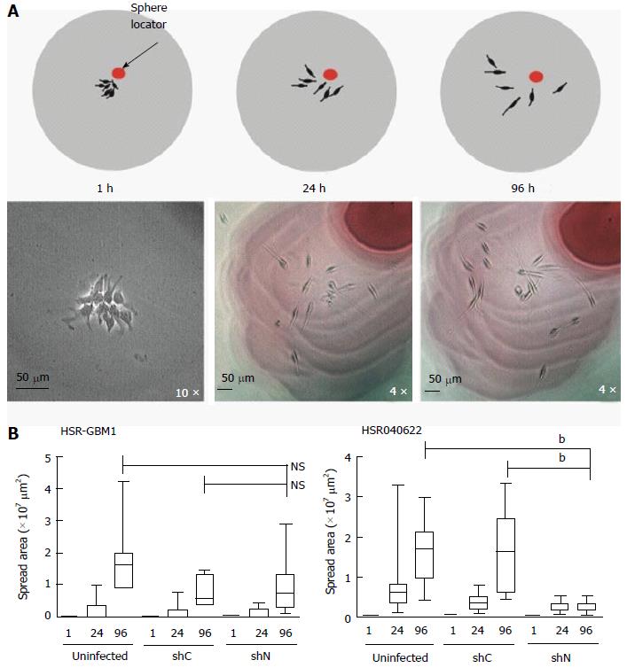
Figure 1 Effect of nestin knockdown on cell migration.
A: Top, cartoon illustrating the experimental design (described in details in materials and methods). Bottom, the two-dimensional area spread of a selected HSR-GBM1 sphere is shown; B: Two dimensional area spread analysis shown no significant difference in cell migration for HSR-GBM1 (left) uninfected, shC, and shN infected cells. In contrast, a significant reduction in cell migration was observed for shN infected HSR040622 cells (right) as compared with uninfected and shC infected cells (NS: Not significant, bP < 0.001; two sided t test). shC: Scrambled control.
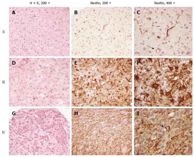
Figure 2 Nestin expression in gliomas.
Diffuse astrocytomas (A) were generally either negative for nestin or contained rare positive cells (B). Endothelial cells were also often nestin positive (C, arrow). Anaplastic astrocytomas (D) generally contained moderate to frequent numbers of strongly nestin positive cells (E, F). Glioblastomas (G) were uniformly nestin positive (H, I).
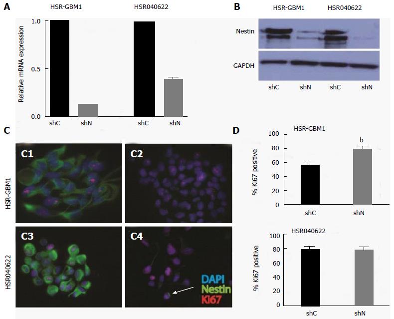
Figure 3 Nestin expression knockdown has no effect on glioblastoma cell proliferation.
Quantitative real time PCR and Western Blot analyses of Nestin mRNA (A) and protein (B) expression in HSR-GBM1 and HSR040622 neurospheres confirming stable knockdown of nestin expression by short hairpin RNA. HSR-GBM1 (C1, C2) and HSR040622 (C3, C4) immuno-staining confirms nestin protein reduction in shNestin (shN) infected cells (C2, C4). Quantification of Ki67 immunoreactivity shown in D (bP < 0.01, two-sided t tests). shC: Scrambled control.
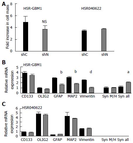
Figure 4 Effect of nestin knockdown on proliferation and the expression of stem cell and differentiation markers.
A: HSR-GBM1 and HSR040622 cells stably expressing the lentivirus driven nestin shRNA show no significant difference in cellular proliferation over a period of 6 d as compared with shC (scrambled control) infected cells; B: In HSR-GBM1, lowered nestin levels has no significant effect on mRNA levels of the neural stem-cell markers CD133 and OLIG2, while statistical significant reduction of mRNA levels of GFAP (glial differentiation), MAP2 (neuronal differentiation), and the intermediate filament Vimentin was observed. The expression of the L-transcript variant of the intermediate filament synemin (syn) was significantly increased; C: In HSR040622, reduced nestin levels had no effect on expression of any of the tested genes mentioned above. It is important to note that the expression of the intermediate filament Synemin was barely detected. (NS = not significant, aP < 0.05, bP < 0.01, dP < 0.001; all two sided t test ). shC: Scrambled control; shN: ShNestin.
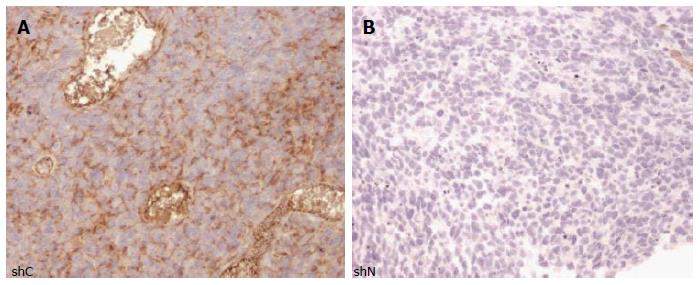
Figure 5 Nestin is not required for xenograft engraftment in HSR-GBM1.
Nestin immunostaining analysis of shC (A) and shN (B) infected HSR-GBM1 cells engrafted into the striatum of athymic nude mice. No significant differences in cellular morphology or migration of tumor cells were apparent at the time the mice were sacrificed. Similar phenotype was observed when uninfected cells were injected (not shown). In all experiments, mice survived for about 63 d post injection. shC: Scrambled control.
- Citation: Lin A, Marchionni L, Sosnowski J, Berman D, Eberhart CG, Bar EE. Role of nestin in glioma invasion. World J Transl Med 2015; 4(3): 78-87
- URL: https://www.wjgnet.com/2220-6132/full/v4/i3/78.htm
- DOI: https://dx.doi.org/10.5528/wjtm.v4.i3.78


