Copyright
©2014 Baishideng Publishing Group Inc.
World J Transl Med. Aug 12, 2014; 3(2): 96-111
Published online Aug 12, 2014. doi: 10.5528/wjtm.v3.i2.96
Published online Aug 12, 2014. doi: 10.5528/wjtm.v3.i2.96
Figure 1 Arsenic methylation pathway in the human body[25].
A: Arsenate reductase or purine nucleoside phosphorylase (PNP); B: Arsenite methyl transferase (As3MT); C: Glutathione S-transferase omega 1 or 2 (GSTO1, GSTO2); D: Arsenite methyl transferase (As3MT). SAHC: S-adenosylhomocysteine; SAM: S-adenosylmethionine; MMAV: Monomethylarsenic acid; MMAIII: Monomethylarsonous acid; DMAV: Dimethylarsenic acid; DMAIII: Dimethylarsinous acid.
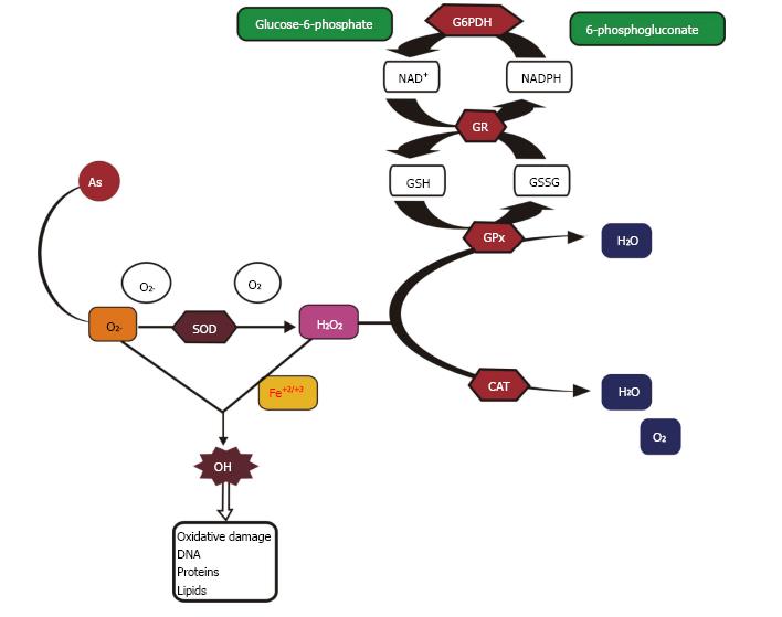
Figure 2 Mechanism of arsenic toxicity: Arsenic enhances the production of superoxide anion radical which results in a higher oxidant level than antioxidant enzymes involved in the detoxification of superoxide anion radical viz.
, superoxide dismutase, catalase, glutathione reductase and glucose-6-phosphate dehydrogenase. SOD: Superoxide dismutase; CAT: Catalase; GPx: Glutathione peroxidase; GR: Glutathione reductase; G-6-PDH: Glucose-6-phosphate dehydrogenase; NAD+: Nicotinamide adenine dinucleotide; NADPH: Nicotinamide adenine dinucleotide phosphate reduced.
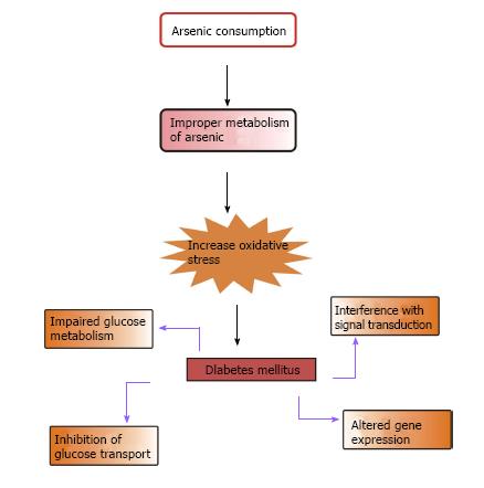
Figure 3 Possible biochemical approaches by which arsenic induces diabetes.
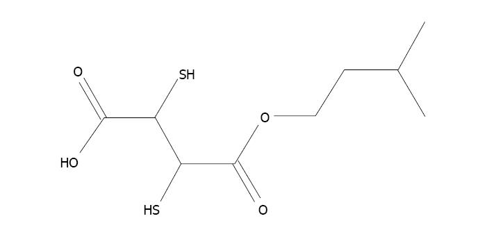
Figure 4 Mono isoamyl 2,3 dimercaptosuccinic acid.
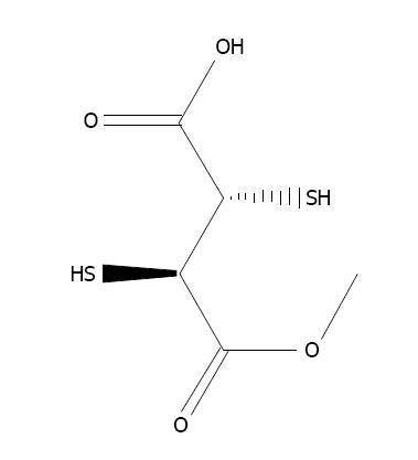
Figure 5 Mono methyl 2,3-dimercaptosuccinic acid.
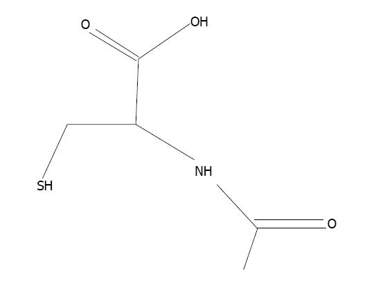
Figure 6 N-acetylcysteine.
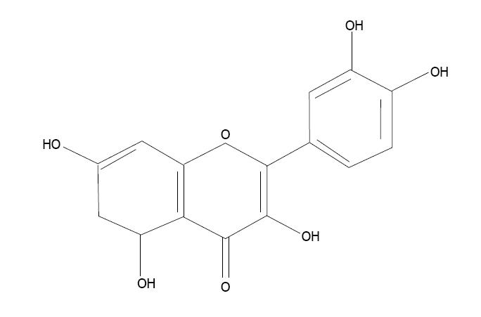
Figure 7 Quercetin.
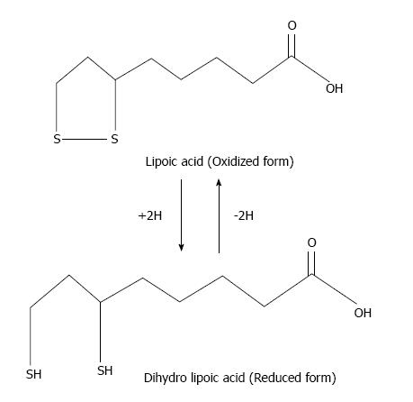
Figure 8 Reduction of lipoic acid.
- Citation: Kulshrestha A, Jarouliya U, Prasad G, Flora S, Bisen PS. Arsenic-induced abnormalities in glucose metabolism: Biochemical basis and potential therapeutic and nutritional interventions. World J Transl Med 2014; 3(2): 96-111
- URL: https://www.wjgnet.com/2220-6132/full/v3/i2/96.htm
- DOI: https://dx.doi.org/10.5528/wjtm.v3.i2.96


