Copyright
©The Author(s) 2023.
World J Transl Med. Aug 31, 2023; 11(1): 1-8
Published online Aug 31, 2023. doi: 10.5528/wjtm.v11.i1.1
Published online Aug 31, 2023. doi: 10.5528/wjtm.v11.i1.1
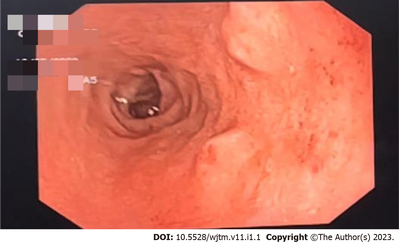
Figure 1 Upper gastrointestinal endoscopy image of a 57-year male with G1 duodenal neuroendocrine tumor.
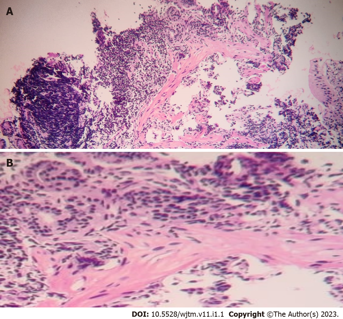
Figure 2 Haematoxylin and eosin staining in low power view of 57-year male with G1 duodenal neuroendocrine tumor.
A: In low power view; B: In high power view.
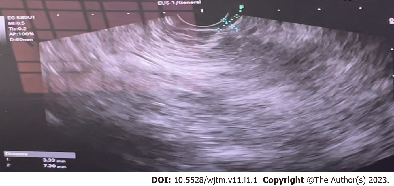
Figure 3 Endoscopic ultrasound image of 57-year male with G1 duodenal neuroendocrine tumor.
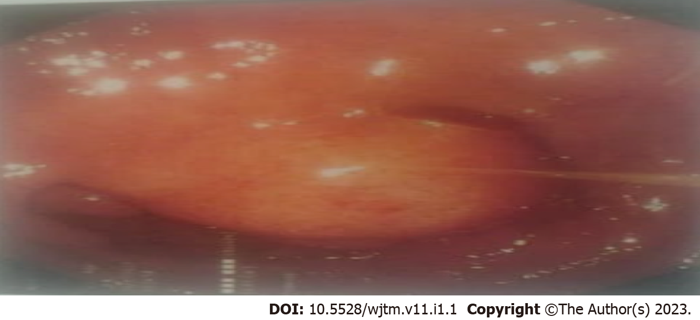
Figure 4 Upper gastrointestinal endoscopy of 52-year male with G1 duodenal neuroendocrine tumor.
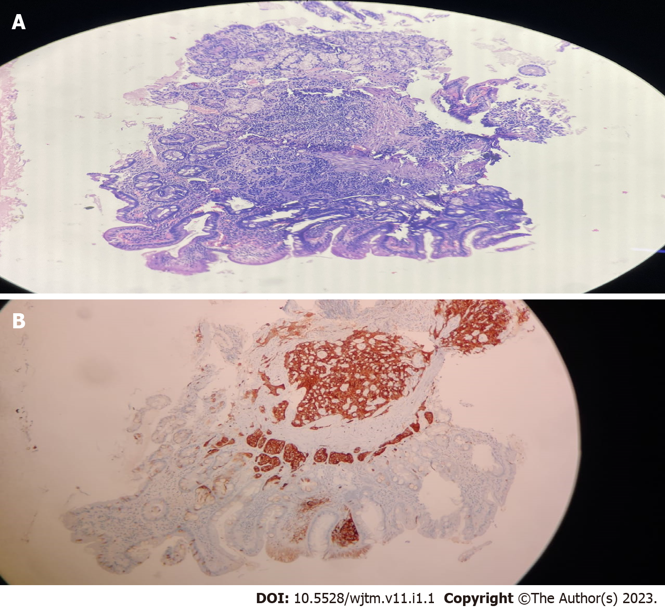
Figure 5 Low power view of a 53-year-old male G1 duodenal neuroendocrine tumor patient.
A: Haematoxylin and eosin staining; B: Immuno
- Citation: Malladi UD, Chimata SK, Bhashyakarla RK, Lingampally SR, Venkannagari VR, Mohammed ZA, Vargiya RV. Duodenal neuroendocrine tumor-tertiary care centre experience: A case report. World J Transl Med 2023; 11(1): 1-8
- URL: https://www.wjgnet.com/2220-6132/full/v11/i1/1.htm
- DOI: https://dx.doi.org/10.5528/wjtm.v11.i1.1


