Copyright
©The Author(s) 2019.
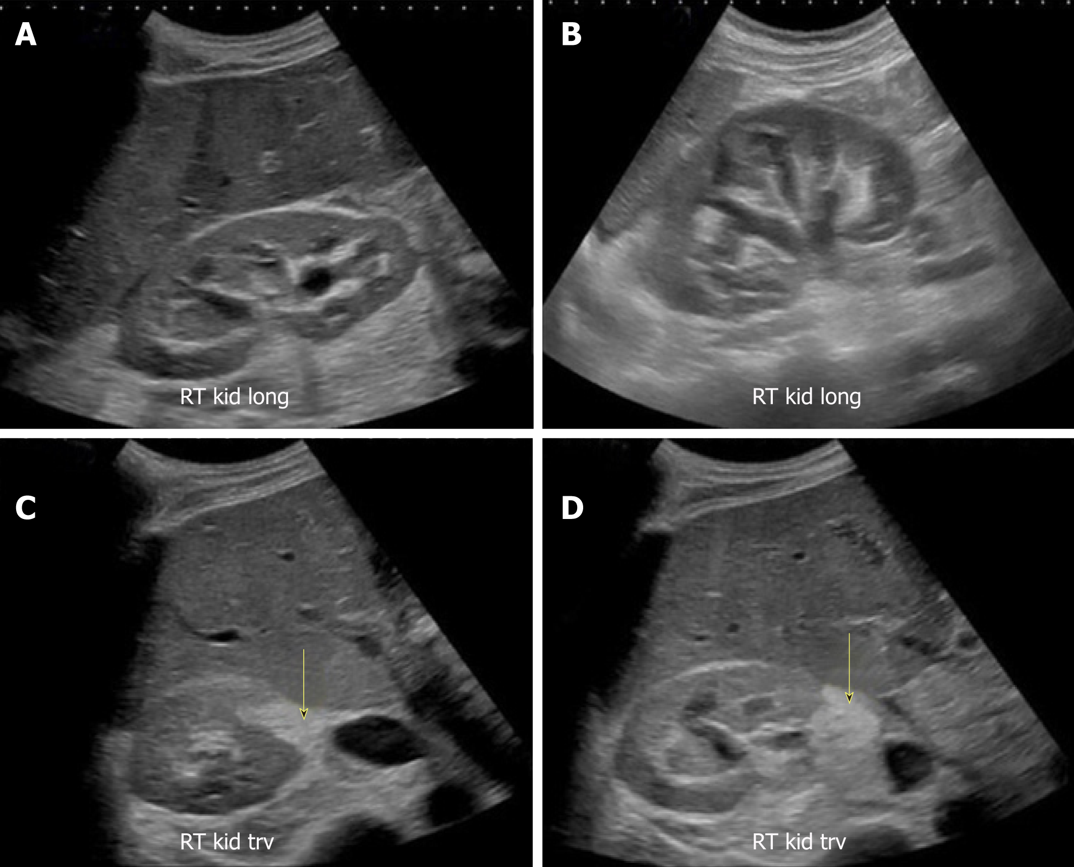
Figure 16 Renal sonogram images.
A, B: Bilateral moderate hydronephrosis and hydroureter; C, D: Transverse views of the right kidney showing echogenic debris (arrows) in the right collecting system in addition to fluid–fluid levels, suggestive of pyonephrosis. Adapted from reference No. 43, first author’s previous work, published under CC BY-NC 4.0 license.
- Citation: Koratala A, Bhattacharya D, Kazory A. Point of care renal ultrasonography for the busy nephrologist: A pictorial review. World J Nephrol 2019; 8(3): 44-58
- URL: https://www.wjgnet.com/2220-6124/full/v8/i3/44.htm
- DOI: https://dx.doi.org/10.5527/wjn.v8.i3.44


