Published online Mar 25, 2025. doi: 10.5527/wjn.v14.i1.96946
Revised: August 10, 2024
Accepted: September 3, 2024
Published online: March 25, 2025
Processing time: 246 Days and 14.5 Hours
Various stone factors can affect the net results of shock wave lithotripsy (SWL). Recently a new factor called variation coefficient of stone density (VCSD) is being considered to have an impact on stone free rates.
To assess the role of VCSD in determining success of SWL in urinary calculi.
Charts review was utilized for collection of data variables. The patients were subjected to SWL, using an electromagnetic lithotripter. Mean stone density (MSD), stone heterogeneity index (SHI), and VCSD were calculated by generating regions of interest on computed tomography (CT) images. Role of these factors were determined by applying the relevant statistical tests for continuous and categorical variables and a P value of < 0.05 was gauged to be statistically signi
There were a total of 407 patients included in the analysis. The mean age of the subjects in this study was 38.89 ± 14.61 years. In total, 165 out of the 407 patients could not achieve stone free status. The successful group had a significantly lower stone volume as compared to the unsuccessful group (P < 0.0001). Skin to stone distance was not dissimilar among the two groups (P = 0.47). MSD was significantly lower in the successful group (P < 0.0001). SHI and VCSD were both significantly higher in the successful group (P < 0.0001).
VCSD, a useful CT based parameter, can be utilized to gauge stone fragility and hence the prediction of SWL outcomes.
Core Tip: In the recent literature various computed tomography (CT) based stone parameters have been studied regarding their role in success of shock wave lithotripsy. These non-contrast CT parameters include skin-stone distance, stone volume, and stone density, which might help in prediction of the success of shock wave lithotripsy (SWL). In recent guidelines, it has been agreed upon that successful outcomes become less likely for harder stones-stone density more than 900-1000 Hounsfield units. Recently, novel parameters such as stone heterogeneity index and variation coefficient of stone density can be representative of stone heterogeneity and can be of utility in prediction of SWL outcome. We, in this study, tried to ascertain their predictive role in shock wave lithotripsy outcomes.
- Citation: Iqbal N, Hasan A, Iqbal S, Noureen S, Akhter S. Role of variation coefficient of stone density in determining success of shock wave lithotripsy in urinary calculi. World J Nephrol 2025; 14(1): 96946
- URL: https://www.wjgnet.com/2220-6124/full/v14/i1/96946.htm
- DOI: https://dx.doi.org/10.5527/wjn.v14.i1.96946
Extracorporeal shock wave lithotripsy (SWL) became an astounding introduction in the armament for treating renal stones. As the time progressed, it precipitously attained popularity across the world. With subtle technological innovations in the techniques of SWL, better procedural outcomes were observed[1]. Owing to this reason, SWL proved itself to have a solid role in the principles of managing kidney and ureter stones[2]. One of the pivotal factors for SWL being an attractive option is its noninvasiveness nature. It can easily be performed as a day case procedure without need for anesthesia[3,4].
In last few years researchers have found that despite the non-invasive nature of SWL procedure, there are certain situations where its effectivity seems to be lower in terms of stone free rates when compared to the outcomes of ureteroscopy (URS), retrograde intrarenal surgery, or percutaneous nephrolithotomy (PCNL). Such procedural failure of SWL might add to prolonged symptoms and burden of ancillary therapy and expenses[5]. Therefore, it entails the fact to identify the predictive tools to predict SWL success and to work out a suitable treatment strategy for patients with upper tract stones.
Stone characteristics, such as locality and size, have been explored in the context of their possible effects on SWL success[6]. Additionally, in the recent past increasing evidence is being collected regarding non-contrast computed tomography (CT) parameters, such as skin-stone distance (SSD), stone volume, and stone density, which might help in prediction of SWL procedural success[7-9]. One of these predictors, SSD, has remained controversial and has failed to be proven as a real predictor[10-12]. Stone density, on the other hand, has proved to be a promising factor in foretelling the SWL outcomes. In recent guidelines it has been agreed upon that successful outcomes become less likely for harder stones, especially when stone density is more than 900-1000 Hounsfield units (HUs). In the latest literature it has been pointed out that some new factors such as stone heterogeneity can influence shock wave outcomes[12-15]. In one study it was found that the inside structural architect of a stone (when seen on CT imaging) helped in predicting extent of stone fragility (in vitro) when subjected to SWL[12-15].
Owing to the absence of any established way of computing stone heterogeneity on CT images, it is imperative to find an authentic and validated form of such calculation. Having said that, it is the need of the hour to dig out CT based tools that might portray stone fragility (heterogeneity) and to look for its capability for predicting SWL outcomes.
Recently it was hypothesized that variation coefficient of stone density (VCSD) can be representative of stone heterogeneity and be of utility in prediction of SWL outcome.
We in this study tried to add and try this novel concept in our clinical practice for treatment of renal and ureter stones. It will not only augment our belief in the authenticity of this newly found concept but also pave a way for its incorpo
In total, 407 patients were included for the final analysis as their CT images were available for the desired computations on PACS system of radiology. Moreover they had completed the follow-up in urology clinic. The inclusion criteria were age more than 18 years (adult age), no prior history of lithotripsy or surgery on ipsilateral side, no anatomical abnormality of the ipsilateral kidney or ureter, and normal coagulation functions. On the other hand, subjects were excluded from study in case of failure to comply with follow-up visits in urology clinic, presence of active urinary tract infection, deranged coagulation profile, and history of prior procedures on the ipsilateral ureter or kidney.
We conducted detailed charts review and prospectively collected data for variables such as patient age, body mass index, gender, stone laterality (left or right), stone anatomical location in the kidney or ureter. This study was approved by the institutional review board of our hospital. On initial visit at urology clinic, all patients were diagnosed after taking their thorough history and performing physical examination prior to subjecting them to SWL. The radiological assess
All patients were subjected to SWL, utilizing the 3rd generation electromagnetic lithotripter Storz Modulith SLX-MX. Patients were positioned supine for procedure. Stone targeting was achieved with utilization of fluoroscopy (Modulith SLX-MX) further assisted with ultrasound (model Aloka SSD-Thousand; 1000). Shock waves were delivered at a rate of 90 shock waves per minute. Initially, 500 shocks were delivered at the energy level 2 and then a gradual ramping up to energy levels 3 and 4 was done for next 2000-2500 shocks. We proclaimed patients in this study to have secured stone free status if there was no proof of residual stone fragments or clinically insignificant residual stone fragments with a size less than 4 mm depicted on plain X-ray (KUB) or abdomen and pelvis ultrasound done three months after the last lithotripsy session. In case of need for ancillary procedures such as PCNL or URS or Double J stenting, subjects were assigned to the SWL procedural failure group.
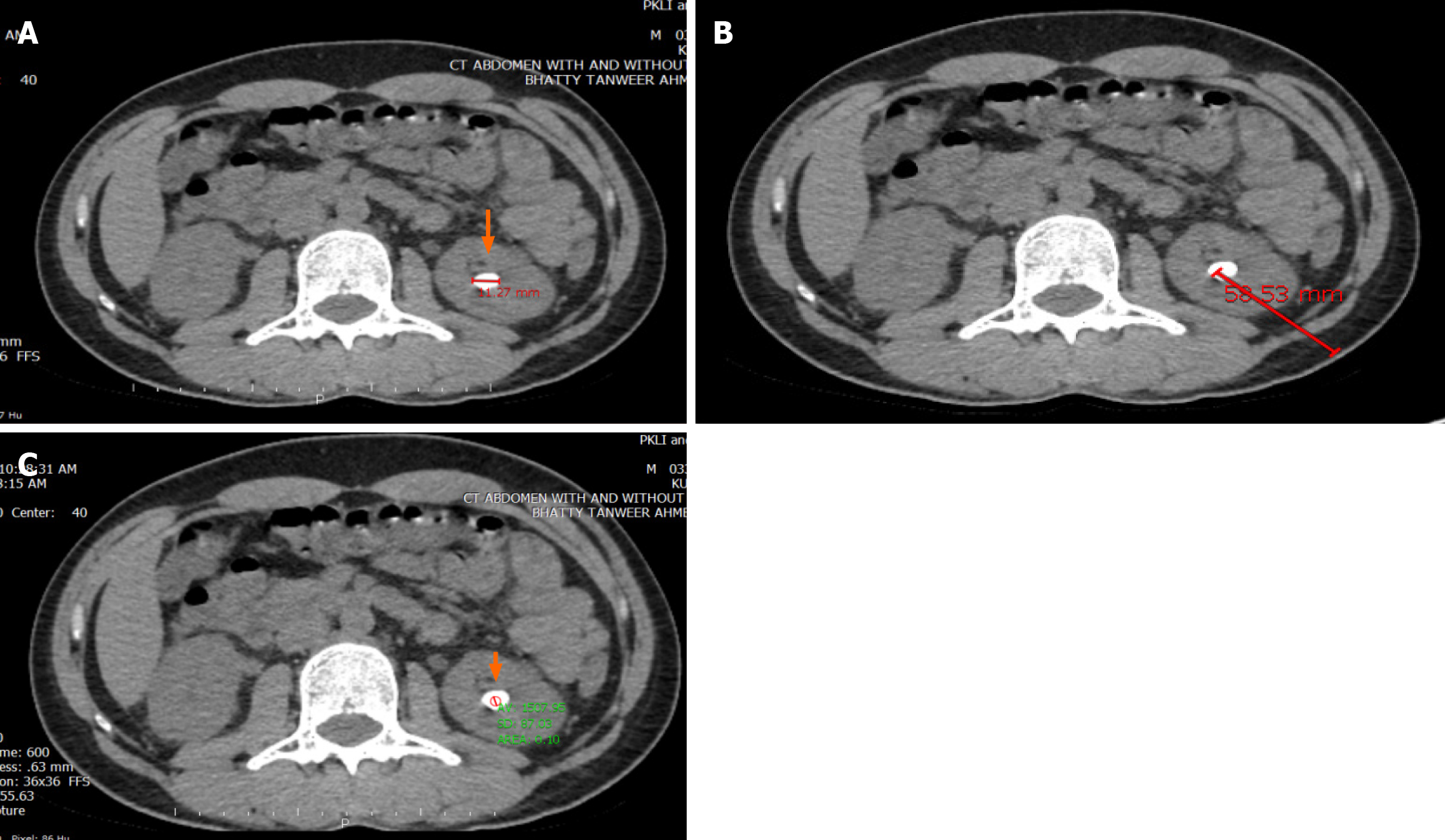
We collected various CT based important variables such as stone density, stone heterogeneity index (SHI), skin-stone distance, stone volume, and VCSD. Two urologists and a radiologist appraised all CT images and utilized the PACS system for this purpose. Stone volume was estimated by using the ellipsoid formula SV = π/6 × (Antero-posterior × Transverse × Cranio-caudal diameters of the stone in mm) and final volume was computed as mm3 (Figure 1A). For computing the SSD values, the methodology illustrated by Pareek et al[15] was used. Skin-stone distance computation is shown in Figure 1B. Computation of mean stone density (MSD; mean value of HUs) was accomplished on an axial image (CT) by generating an elliptical region of interest, portraying stone in its longest dimension. Particular attention was given not to include any soft tissue while gauging the stone density (Figure 1C).
Mean stone density (MSD) is expressed as the mean value of HUs computed in the region of interest. SHI was calculated as the standard deviation value of HUs in that same designated region of interest as shown in Figure 1. VCSD, the variable of pivotal interest in the present study, was interpreted and computed by utilizing the formula [stone density standard deviation (SDSD)/MSD × 100%]. Please refer to Figure 1 for understanding how stone volume, SSD, and MSD, SDSD (SHI), VCSD were calculated.
After gathering of information for the variables in specified proformas, it was entered in Statistical Package for Social Sciences, version 16 (SPSS Inc.; Chicago, IL, United States) for computations and statistical analyses. Continuous variables such as calculus volume, subject age, stone density, and SHI were compared by employing the Student’s t-test and Mann Whitney test where applicable. Categorical variables were compared by using the Pearson’s χ2 test. A P value of < 0.05 (Two-tailed) was ascertained to be statistically significant while constructing these comparisons.
There were a total of 407 patients included in the analysis. Among these, 113 were female patients. The mean age of the subjects in this study was 38.89 ± 14.61 years (Table 1). Most of the stones were located on the left side (Table 1). Median stone density was 900 HUs. In 316 of the patients stones were lying in renal location (Table 1).
| Demographic parameter | Result |
| Age (years) | 38.89 ± 14.61 |
| Gender, n (%) | |
| Male | 294 (72.2) |
| Female | 113 (27.8) |
| Stone volume | 223.34 ± 77.01 |
| SSD (cm)1 | 9.52 ± 2.47 |
| Stone density (HUs) | 900 (110-1570) |
| SHI (HUs)2 | 174.85 (17.7-583.9) |
| VCSD3 | 22 (3-83) |
| Stone laterality, n (%) | |
| Left sided | 213 (52.3) |
| Right sided | 194 (47.7) |
| Location stone, n (%) | |
| Renal | 316 (77.64) |
| Ureter | 91 (22.36) |
In total, 165 out of the 407 patients could not achieve stone free status. Comparison of different patient related characteristics and stone features between the successful and unsuccessful groups is summarized in Table 2. It is evident from Table 2 that there was no significant difference in terms of age (P = 0.93) or gender ratio (P = 0.78) between the successful and unsuccessful groups of patients. The successful group (group 2) had a significantly lower stone volume as compared to the group 1 (the unsuccessful group) (P < 0.0001). SSD was not significantly different between the two groups (P = 0.47). Moreover, MSD, SHI, and VCSD were significantly different between the two groups. MSD was significantly lower in group 2 (the successful group) (P < 0.0001). SHI and VCSD were both significantly higher in the successful group (P < 0.0001).
| Parameter | Unsuccessful group | Successful group | P value |
| Age (years)1 | 38.82 ± 13.84 | 38.94 ± 15.13 | 0.93 |
| Gender, n (%) | 0.78 | ||
| Male | 118 | 176 | |
| Female | 47 | 66 | |
| Stone volume2 | 240 (80-439) | 200 (36-400) | < 0.0001 |
| SSD (cm)1 | 9.63 ± 2.32 | 9.45 ± 2.56 | 0.47 |
| Stone density (HUs)2 | 1217.30 (318-2000) | 720.00 (110-1800) | 0.0001 |
| SHI (HUs)2 | 172.95 (22.7-571.4) | 190.15 (17.7-583.9) | < 0.0001 |
| VCSD2 | 15 (2-92) | 28 (4-83) | < 0.0001 |
| Stone laterality, n (%)) | 0.19 | ||
| Left sided | 80 | 133 | |
| Right sided | 85 | 109 | |
| Stone location | 0.69 | ||
| Upper pole | 12 | 17 | |
| Mid pole | 29 | 38 | |
| Lower pole | 46 | 79 | |
| Pelvis | 39 | 55 | |
| Ureter | 39 | 53 | |
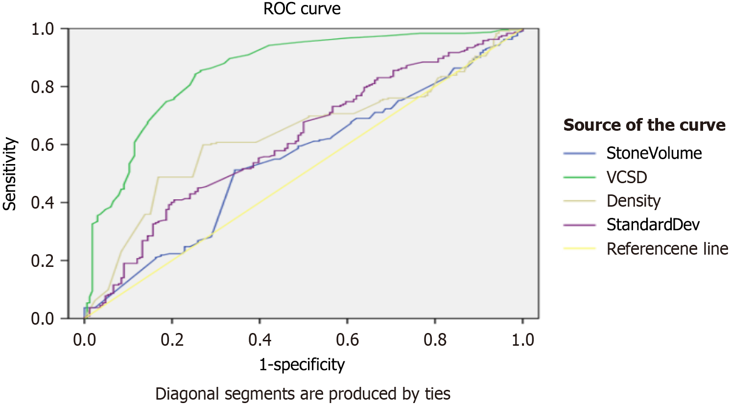
Table 3 summarizes the net results after application of univariate and multivariate logistic regression for variables that could predict success of SWL after first session for subjects included in this study. In univariate analysis, higher MSD (P < 0.0001), lower values of standard deviation of stone density (P < 0.0001), lower values of VCSD (P < 0.0001), and larger stone volume (P < 0.0001) were linked to failure of the SWL procedure. On computing multivariate analysis, higher MSD (P < 0.0001), lower values of VCSD (P < 0.0001), and larger stone volume (P < 0.005) were strong predictors of SWL failure (Table 3). Figure 2 shows the receiver operating characteristic (ROC) curves generated for VCSD, SHI, stone volume, and stone density, which suggest good predictive value of VCSD for SWL success rates.
| Variable | Univariate | Multivariate | ||||
| OR | 95%CI | P value | OR | 95%CI | P value | |
| Mean stone density | 1.003 | 1.003-1.004 | < 0.0001 | 1.003 | 0.424-1.346 | < 0.0001 |
| Standard deviation of stone density | 0.996 | 0.995-0.998 | < 0.0001 | 0.997 | 0.993-1.001 | 0.13 |
| VCSD (%)1 | 0.895 | 0.874-0.917 | < 0.0001 | 0.929 | 0.894-0.964 | < 0.0001 |
| Stone volume (mm3) | 1.009 | 1.007-1.012 | < 0.0001 | 1.005 | 1.002-1.009 | < 0.005 |
| Age (year) | 0.99 | 0.985-1.012 | 0.86 | |||
| Skin-stone distance | 1.02 | 0.947-1.112 | 0.52 | |||
| Gender (male) | 0.949 | 0.611-1.475 | 0.81 | |||
Research studies in last few years regarding outcomes of SWL have demonstrated considerable variations of success rates (as miniscule as 32% up to 95%). Such variations in outcomes forced urologists and researchers to hunt for factors that might influence the main outcome and the decision-making process by the treating urologist[16,17]. An unprecedented study was reported in 2005, which narrated the concept that SSD calculated by using NCCT can be a crucial predictive factor with regards to SWL procedural success in patients. It was the introductory study to correlate outcomes of ESWL to SSD[12].
Next studies led by other authors revealed effects of stone size and stone density upon net results of SWL[18,19]. As more findings accumulated in favor of role of these factors, step by step efforts were done to find more factors that could predict post-procedural net results. These efforts were done to aid in more suitable selection of patients to be subjected to SWL.
Recently some more urinary stone features (based on CT images) have been investigated to look for their possible effect on net results of the SWL procedures. The present study examined these new radiologic features of stones seen on CT images, including the heterogeneity of stone density and VCSD and obtained some important observations. To the best of our knowledge, this is the first study where the confounding factor of SSD was eliminated between the stone free and stone failure groups due to the type of patients included.
As described already, the role of stone density has been established over various reports in past few years. It has become a beneficial parameter to gauge and foretell SWL success[16-19]. It is therefore utilized widely nowadays in urological clinics regarding counselling patients about possible outcomes. In a study by Bulut et al[19] differences in stone characteristics, including stone density and volume, were statistically significant in patients who achieved stone-free status by SWL or not. Nevertheless, MSD can depict just the average value of hardness of the stone. In other words, the internal structural and chemical diversity of a urinary stone (the heterogeneity of chemical composition of stone) is not given consideration. Recently limited studies have tried to look for feasible role of heterogeneity of stones as vital feature for foretelling success of SWL[17-20]. These studies mentioned the possibility of usefulness of the SHI (on the basis of CT images). Recently, Lee et al[20] suggested that SHI could be used to predict net result of SWL in subjects with urinary calculi. However, it is pertinent to note here that SHI illustrates standard deviation values of the mean value of stone density, in that case it is likely to be governed by the mean value, an important point to ponder over. According to Lee et al[20], in subjects who had higher stone density (MSD ≥ 1000 HUs), the SHI value was strikingly higher in the one-session success group compared to the failure group (308 ± 92 HU vs 251 ± 55 HU). It underscored the fact that SHI is an important predictor of SWL success rates.
On the contrary, the new entry of a tool called variation coefficient, which can be obtained by dividing the value of the standard deviation of stone density by the mean value of stone density, can be a more useful predictive tool. Yamashita et al[21] postulated that VCSD might represent stone heterogeneity better as compared to SHI only. Yamashita et al[21] advocated VCSD as a new tool that may be utilized for predicting SWL net results. They mentioned that high VCSD values basically show substantial dispersion of stone density (so it can give comprehensible picture of heterogeneity in the inner structure of the stone in question). By performing ROC curve analysis, they deduced a cut off value of 51.3% (for VCSD) for successful SWL. They detected that success for procedure was nearly 64% for a value of VCSD higher than 51.3%. On the other hand, in stones having a less than 51% value of VCSD, only 26% of the study subjects achieved stone free status. Previous reports have mentioned the role of heterogeneity of stones to be of vital significance. The more the heterogeneity of stone density, the more the fragility of the stone and such a stone can be easily cracked when subjected to SWL. The VCSD values are higher in stones having higher values of stone heterogeneity. A good point about VCSD is that it has more practicable value as it gives the ratio of the heterogeneity of stones to the true mean density values of stones. Thus, two stones may have the same heterogeneity values but if their mean density values are different, then in such tricky scenario VCSD value will guide better regarding the inclination of a stone to fragility. For instance, if heterogeneity values of two different stones are 300 (similar) and MSD values are 500 and 1000 HUs (different for the two stones), VCSD shall be a better guiding tool because for first stone VCSD = 61% (higher value) and for second stone VCSD = 30% (lower value). The stone with higher VCSD indicates a higher degree of dispersion of density values and is thus affected not only by heterogeneity value but also by the MSD value at the same time[20,21].
Yamashita et al[21] found that higher VCSD is the independent significant predictor of SWL success (P < 0.001) in overall patients. We also had similar results as can be seen in Table 3. Median stone density, median SDSD, and median VCSD were 545.0 HUs, 283.0 HUs, and 54.0%, respectively, in the study by Yamashita et al[21]. The present study was slightly different from their study in a way that we had considered impact of VCSD in stones having higher mean density (median 900 HUs) with lower SDSD (median 174.85) and VCSD values (22%) as compared to theirs (Table 1). We had a stone free rate of 59.4% as compared to their success rate of 47.7%. This may be due to their higher SSD (10.4 cm) as compared to that of the present study (9.52 cm). Second, they considered patients with multiple stones that could have resulted inferior success rates. Third, they had comparatively larger median stone volume resulting in higher rates of failure.
SHI and VCSD are new stone CT-based parameters being investigated nowadays. Prior to this, stone density, SSD, and stone volume have been explored regarding their effect on SWL success rates. Ouzaid et al[22] inferred that stones of more than 970 HUs had higher chances of SWL failure. El-Nahas et al[10] pointed out that an MSD value of more than 1000 HUs was a vital tool to predict SWL procedural failure.
Ozgor F et al[23] in their remarkable research work mentioned that differences in stone characteristics, including stone location, density, and volume, were statistically significant in patients who achieved stone-free status by SWL or not (P < 0.001, P < 0.001, and P < 0.001, respectively). In another similar study, stone free status was attained in 160 patients (57.7%), and 117 (42.3%) patients were labeled to have failed the procedure. Differences between these two groups in terms of stone volume, stone density, and SSD were significant[24].
The present study has some strengths, e.g., the SSD was lesser as compared to contemporary studies to remove its confounding effect that could have affected the results differently. However, this study also has limitations such as lack of randomization based on different stone size and stone density. Lastly, it was a single center study, so its results may not necessarily be generalized. There are no multicenter studies regarding this aspect of stone feature. We believe that more investigative studies are required to ascertain the role of VCSD concept in decision pathway utilized by urologists. It can be a useful tool and can be informative for both physicians and patients for making a shared decision regarding treatment planning.
It is inferred that VCSD is an advantageous CT-based tool to predict stone success rate. Moreover, it can be of help to both the physicians and patients in making shared decisions regarding SWL procedure. Incorporation of VCSD in nomograms may further enhance its role in such decision-making.
We are thankful to Prof. Saeed Akhter for his unwavering support.
| 1. | Chaussy C, Brendel W, Schmiedt E. Extracorporeally induced destruction of kidney stones by shock waves. Lancet. 1980;2:1265-1268. [RCA] [PubMed] [DOI] [Full Text] [Cited by in Crossref: 826] [Cited by in RCA: 706] [Article Influence: 15.7] [Reference Citation Analysis (0)] |
| 2. | Türk C, Petřík A, Sarica K, Seitz C, Skolarikos A, Straub M, Knoll T. EAU Guidelines on Interventional Treatment for Urolithiasis. Eur Urol. 2016;69:475-482. [RCA] [PubMed] [DOI] [Full Text] [Cited by in Crossref: 775] [Cited by in RCA: 1090] [Article Influence: 109.0] [Reference Citation Analysis (0)] |
| 3. | Iqbal N, Malik Y, Nadeem U, Khalid M, Pirzada A, Majeed M, Malik HA, Akhter S. Comparison of ureteroscopic pneumatic lithotripsy and extracorporeal shock wave lithotripsy for the management of proximal ureteral stones: A single center experience. Turk J Urol. 2018;44:221-227. [RCA] [PubMed] [DOI] [Full Text] [Cited by in Crossref: 10] [Cited by in RCA: 13] [Article Influence: 1.9] [Reference Citation Analysis (0)] |
| 4. | Yamashita S, Kohjimoto Y, Iwahashi Y, Iguchi T, Nishizawa S, Kikkawa K, Hara I. Noncontrast Computed Tomography Parameters for Predicting Shock Wave Lithotripsy Outcome in Upper Urinary Tract Stone Cases. Biomed Res Int. 2018;2018:9253952. [RCA] [PubMed] [DOI] [Full Text] [Full Text (PDF)] [Cited by in Crossref: 7] [Cited by in RCA: 19] [Article Influence: 2.7] [Reference Citation Analysis (0)] |
| 5. | Wolf JS Jr. Treatment selection and outcomes: ureteral calculi. Urol Clin North Am. 2007;34:421-430. [RCA] [PubMed] [DOI] [Full Text] [Cited by in Crossref: 77] [Cited by in RCA: 83] [Article Influence: 4.6] [Reference Citation Analysis (0)] |
| 6. | Kanao K, Nakashima J, Nakagawa K, Asakura H, Miyajima A, Oya M, Ohigashi T, Murai M. Preoperative nomograms for predicting stone-free rate after extracorporeal shock wave lithotripsy. J Urol. 2006;176:1453-6; discussion 1456. [RCA] [PubMed] [DOI] [Full Text] [Cited by in Crossref: 99] [Cited by in RCA: 99] [Article Influence: 5.2] [Reference Citation Analysis (0)] |
| 7. | Wiesenthal JD, Ghiculete D, Ray AA, Honey RJ, Pace KT. A clinical nomogram to predict the successful shock wave lithotripsy of renal and ureteral calculi. J Urol. 2011;186:556-562. [RCA] [PubMed] [DOI] [Full Text] [Cited by in Crossref: 67] [Cited by in RCA: 66] [Article Influence: 4.7] [Reference Citation Analysis (0)] |
| 8. | Tanaka M, Yokota E, Toyonaga Y, Shimizu F, Ishii Y, Fujime M, Horie S. Stone attenuation value and cross-sectional area on computed tomography predict the success of shock wave lithotripsy. Korean J Urol. 2013;54:454-459. [RCA] [PubMed] [DOI] [Full Text] [Full Text (PDF)] [Cited by in Crossref: 23] [Cited by in RCA: 32] [Article Influence: 2.7] [Reference Citation Analysis (0)] |
| 9. | Choi JW, Song PH, Kim HT. Predictive factors of the outcome of extracorporeal shockwave lithotripsy for ureteral stones. Korean J Urol. 2012;53:424-430. [RCA] [PubMed] [DOI] [Full Text] [Full Text (PDF)] [Cited by in Crossref: 26] [Cited by in RCA: 42] [Article Influence: 3.2] [Reference Citation Analysis (0)] |
| 10. | El-Nahas AR, El-Assmy AM, Mansour O, Sheir KZ. A prospective multivariate analysis of factors predicting stone disintegration by extracorporeal shock wave lithotripsy: the value of high-resolution noncontrast computed tomography. Eur Urol. 2007;51:1688-93; discussion 1693. [RCA] [PubMed] [DOI] [Full Text] [Cited by in Crossref: 211] [Cited by in RCA: 219] [Article Influence: 12.2] [Reference Citation Analysis (0)] |
| 11. | Bandi G, Meiners RJ, Pickhardt PJ, Nakada SY. Stone measurement by volumetric three-dimensional computed tomography for predicting the outcome after extracorporeal shock wave lithotripsy. BJU Int. 2009;103:524-528. [RCA] [PubMed] [DOI] [Full Text] [Cited by in Crossref: 81] [Cited by in RCA: 96] [Article Influence: 5.6] [Reference Citation Analysis (0)] |
| 12. | Kim SC, Burns EK, Lingeman JE, Paterson RF, McAteer JA, Williams JC Jr. Cystine calculi: correlation of CT-visible structure, CT number, and stone morphology with fragmentation by shock wave lithotripsy. Urol Res. 2007;35:319-324. [RCA] [PubMed] [DOI] [Full Text] [Cited by in Crossref: 60] [Cited by in RCA: 53] [Article Influence: 2.9] [Reference Citation Analysis (0)] |
| 13. | Haas F. Response to comments by Ervin G. Erdös. FASEB J. 2008;22:1623-1624. [RCA] [DOI] [Full Text] [Reference Citation Analysis (0)] |
| 14. | Zarse CA, Hameed TA, Jackson ME, Pishchalnikov YA, Lingeman JE, McAteer JA, Williams JC Jr. CT visible internal stone structure, but not Hounsfield unit value, of calcium oxalate monohydrate (COM) calculi predicts lithotripsy fragility in vitro. Urol Res. 2007;35:201-206. [RCA] [PubMed] [DOI] [Full Text] [Full Text (PDF)] [Cited by in Crossref: 80] [Cited by in RCA: 66] [Article Influence: 3.7] [Reference Citation Analysis (0)] |
| 15. | Pareek G, Hedican SP, Lee FT Jr, Nakada SY. Shock wave lithotripsy success determined by skin-to-stone distance on computed tomography. Urology. 2005;66:941-944. [RCA] [PubMed] [DOI] [Full Text] [Cited by in Crossref: 188] [Cited by in RCA: 197] [Article Influence: 9.9] [Reference Citation Analysis (0)] |
| 16. | Dogan HS, Altan M, Citamak B, Bozaci AC, Karabulut E, Tekgul S. A new nomogram for prediction of outcome of pediatric shock-wave lithotripsy. J Pediatr Urol. 2015;11:84.e1-84.e6. [RCA] [PubMed] [DOI] [Full Text] [Cited by in Crossref: 47] [Cited by in RCA: 50] [Article Influence: 5.0] [Reference Citation Analysis (0)] |
| 17. | Ghoneim IA, Ziada AM, Elkatib SE. Predictive factors of lower calyceal stone clearance after Extracorporeal Shockwave Lithotripsy (ESWL): a focus on the infundibulopelvic anatomy. Eur Urol. 2005;48:296-302; discussion 302. [RCA] [PubMed] [DOI] [Full Text] [Cited by in Crossref: 46] [Cited by in RCA: 55] [Article Influence: 2.8] [Reference Citation Analysis (0)] |
| 18. | Park BH, Choi H, Kim JB, Chang YS. Analyzing the effect of distance from skin to stone by computed tomography scan on the extracorporeal shock wave lithotripsy stone-free rate of renal stones. Korean J Urol. 2012;53:40-43. [RCA] [PubMed] [DOI] [Full Text] [Full Text (PDF)] [Cited by in Crossref: 23] [Cited by in RCA: 31] [Article Influence: 2.4] [Reference Citation Analysis (0)] |
| 19. | Bulut M, Dinçer E, Coşkun A, Can U, Telli O. Is Triple D Score Effective to Predict the Stone-Free Rate After Shockwave Lithotripsy in Pediatric Population? J Endourol. 2023;37:207-211. [RCA] [PubMed] [DOI] [Full Text] [Reference Citation Analysis (0)] |
| 20. | Lee JY, Kim JH, Kang DH, Chung DY, Lee DH, Do Jung H, Kwon JK, Cho KS. Stone heterogeneity index as the standard deviation of Hounsfield units: A novel predictor for shock-wave lithotripsy outcomes in ureter calculi. Sci Rep. 2016;6:23988. [RCA] [PubMed] [DOI] [Full Text] [Full Text (PDF)] [Cited by in Crossref: 36] [Cited by in RCA: 49] [Article Influence: 5.4] [Reference Citation Analysis (0)] |
| 21. | Yamashita S, Kohjimoto Y, Iguchi T, Nishizawa S, Iba A, Kikkawa K, Hara I. Variation Coefficient of Stone Density: A Novel Predictor of the Outcome of Extracorporeal Shockwave Lithotripsy. J Endourol. 2017;31:384-390. [RCA] [PubMed] [DOI] [Full Text] [Cited by in Crossref: 19] [Cited by in RCA: 31] [Article Influence: 3.9] [Reference Citation Analysis (0)] |
| 22. | Ouzaid I, Al-qahtani S, Dominique S, Hupertan V, Fernandez P, Hermieu JF, Delmas V, Ravery V. A 970 Hounsfield units (HU) threshold of kidney stone density on non-contrast computed tomography (NCCT) improves patients' selection for extracorporeal shockwave lithotripsy (ESWL): evidence from a prospective study. BJU Int. 2012;110:E438-E442. [RCA] [PubMed] [DOI] [Full Text] [Cited by in Crossref: 92] [Cited by in RCA: 104] [Article Influence: 8.0] [Reference Citation Analysis (0)] |
| 23. | Ozgor F, Tosun M, Kayali Y, Savun M, Binbay M, Tepeler A. External Validation and Evaluation of Reliability and Validity of the Triple D Score to Predict Stone-Free Status After Extracorporeal Shockwave Lithotripsy. J Endourol. 2017;31:169-173. [RCA] [PubMed] [DOI] [Full Text] [Cited by in Crossref: 8] [Cited by in RCA: 15] [Article Influence: 1.9] [Reference Citation Analysis (0)] |
| 24. | Iqbal N, Hasan A, Singh G, Hassan MH, Nazar A, Khilan MH, Malik SI, Khawaja MA, Akhter S, Iqbal D, Khan F. Use Of Computed Tomography-Based Nomogram In Adult Age Patients To Predict Success Rates After Shock Wave Lithotripsy For Renal Stones: A Single Center Experience. J Ayub Med Coll Abbottabad. 2021;33:386-392. [PubMed] |


