Copyright
©The Author(s) 2018.
Figure 1 The results of light microscopy.
A and B: The glomerulus shows epithelial hypercellularity at the tubular pole, where a confluence of the tubular cells at the tubular outlet is observed (arrows). No collapse of the glomerular tufts associated with human immunodeficiency virus-associated nephropathy (HIVAN) is seen (A: Periodic acid-Schiff stain: Original magnification 400 ×; B: Periodic acid-methenamine-silver-HE stain: Original magnification 400 ×); C: Diffuse tubular atrophy and interstitial fibrosis are seen. However, infiltration of the inflammatory cells is sparse and microcystic tubular dilatation associated with HIVAN is absent (Masson’s trichrome stain: Original magnification 100 ×).
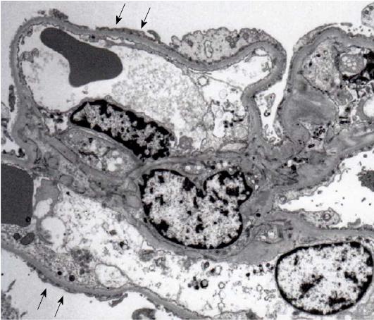
Figure 2 The results of electron microscopy.
Electron microscopy reveals a wide range of foot process effacement (arrows). No tubulo-reticular inclusions in the glomerular endothelium are seen and electron-dense deposits are absent (original magnification: 3000 ×).
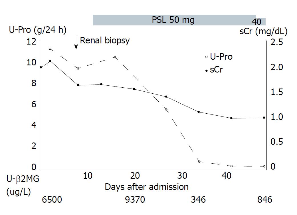
Figure 3 Clinical course after admission.
The solid line with closed circles indicates serum creatinine (sCr) levels, and the dashed line with open circles indicates daily urinary protein (U-Pro) excretion levels. PSL: Prednisolone, U-β2MG; Urinary β2-microglobulin.
- Citation: Goto D, Ohashi N, Takeda A, Fujigaki Y, Shimizu A, Yasuda H, Ohishi K. Case of human immunodeficiency virus infection presenting as a tip variant of focal segmental glomerulosclerosis: A case report and review of the literature. World J Nephrol 2018; 7(4): 90-95
- URL: https://www.wjgnet.com/2220-6124/full/v7/i4/90.htm
- DOI: https://dx.doi.org/10.5527/wjn.v7.i4.90


