Copyright
©The Author(s) 2017.
World J Nephrol. May 6, 2017; 6(3): 100-110
Published online May 6, 2017. doi: 10.5527/wjn.v6.i3.100
Published online May 6, 2017. doi: 10.5527/wjn.v6.i3.100
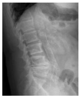
Figure 1 Abdominal aortic calcification seen on lateral lumbar spine radiograph.
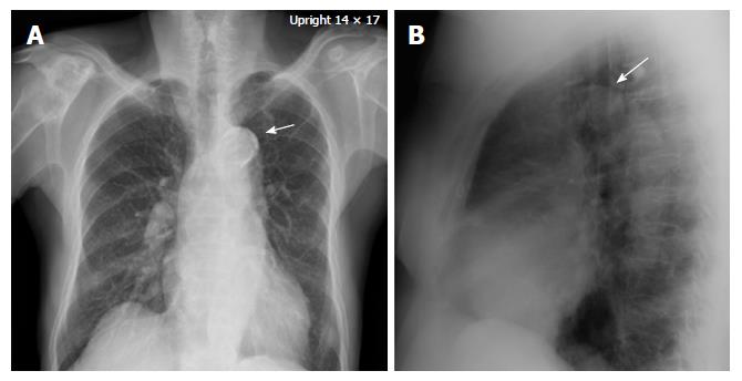
Figure 2 Aortic arch calcification (arrow) seen on postero-anterior chest radiograph (A) and lateral chest radiograph (B).
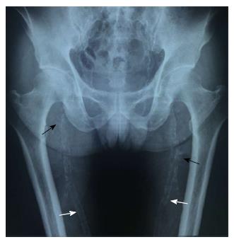
Figure 3 Plaque-like intimal calcification (black arrow) and uniform linear railroad track-like medial calcification (white arrow).
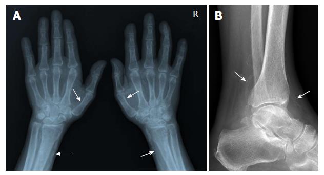
Figure 4 Medial calcification in small arteries in hands (A) and foot (B).
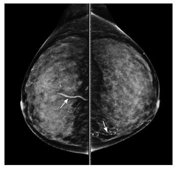
Figure 5 Breast arterial calcification with the typical linear tram-track medial-type calcification (arrows).
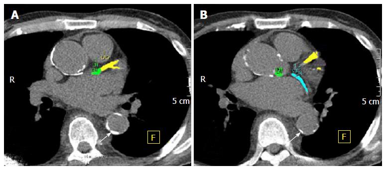
Figure 6 Coronary artery calcification (marked with colors) and descending thoracic aortic calcification (arrows) seen on non-contrast multislice computed tomography.
- Citation: Disthabanchong S, Boongird S. Role of different imaging modalities of vascular calcification in predicting outcomes in chronic kidney disease. World J Nephrol 2017; 6(3): 100-110
- URL: https://www.wjgnet.com/2220-6124/full/v6/i3/100.htm
- DOI: https://dx.doi.org/10.5527/wjn.v6.i3.100


