Copyright
©The Author(s) 2016.
World J Nephrol. Sep 6, 2016; 5(5): 418-428
Published online Sep 6, 2016. doi: 10.5527/wjn.v5.i5.418
Published online Sep 6, 2016. doi: 10.5527/wjn.v5.i5.418
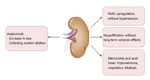
Figure 1 Physiological renal changes during pregnancy.
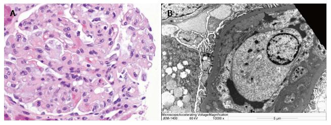
Figure 2 Light (A) and electron (B) microscopy.
A: Glomeruli with capillary loops occluded by swollen endothelial cells, e.g., endotheliosis; B: Capillary loop with subendothelial widening by flocculent material and occlusion by swollen endothelial cells. Courtesy of Naima Carter-Monroe.
- Citation: Berry C, Atta MG. Hypertensive disorders in pregnancy. World J Nephrol 2016; 5(5): 418-428
- URL: https://www.wjgnet.com/2220-6124/full/v5/i5/418.htm
- DOI: https://dx.doi.org/10.5527/wjn.v5.i5.418


