Copyright
©The Author(s) 2016.
World J Nephrol. May 6, 2016; 5(3): 288-299
Published online May 6, 2016. doi: 10.5527/wjn.v5.i3.288
Published online May 6, 2016. doi: 10.5527/wjn.v5.i3.288
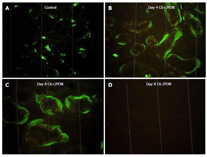
Figure 1 The tubulointerstitial deposition of C5b-9 is increased in C6+ rats with protein-overload nephropathy.
Representative sections are shown. A: Animal injected with saline (control); B: C6+ animal with PON on day 4; C: C6+ animal with PON on day 8; D: C6- animal with PON on day 8 (× 400 magnification). Predominant basolateral deposition of C5b-9 was present in PON. PON: Protein-overload nephropathy.
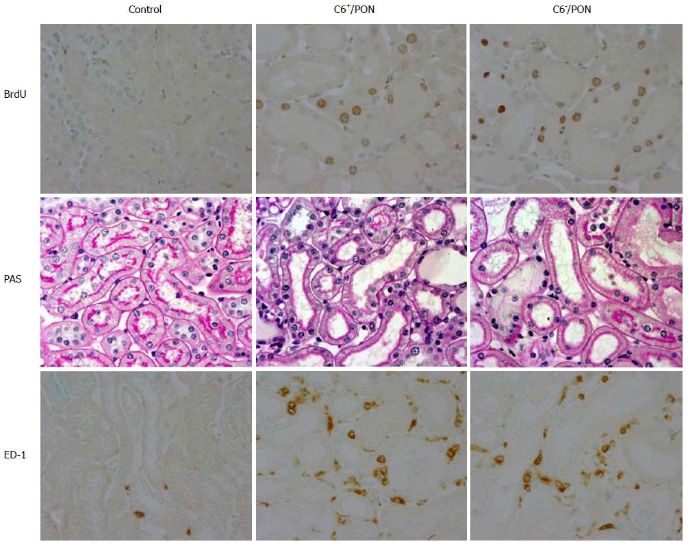
Figure 2 Cortical tubulointerstitial injury in control and protein-overload nephropathy groups on day 8.
Representative renal cortical sections (magnification × 400) show immunostaining for BrdU, periodic acid-schiff and ED-1.
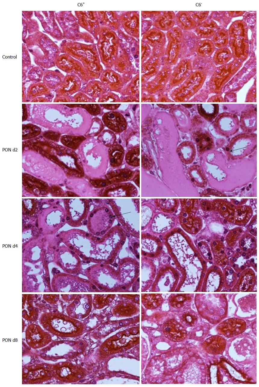
Figure 3 The renal cortical expression of proliferating cell nuclear antigen is increased in protein-overload nephropathy.
Representative sections (× 200 magnification) are shown. PCNA positive TEC nuclei are labeled purple (Nickel-enhanced DAB-positive, arrows). Brush border are labeled with anti-Fx1A (brown) and counterstained with periodic acid-schiff. PCNA: Proliferating cell nuclear antigen; TEC: Tubular epithelial cells.
- Citation: Rangan GK. C5b-9 does not mediate tubulointerstitial injury in experimental acute glomerular disease characterized by selective proteinuria. World J Nephrol 2016; 5(3): 288-299
- URL: https://www.wjgnet.com/2220-6124/full/v5/i3/288.htm
- DOI: https://dx.doi.org/10.5527/wjn.v5.i3.288


