Copyright
©2016 Baishideng Publishing Group Co.
World J Nephrol. Mar 6, 2016; 5(2): 125-138
Published online Mar 6, 2016. doi: 10.5527/wjn.v5.i2.125
Published online Mar 6, 2016. doi: 10.5527/wjn.v5.i2.125
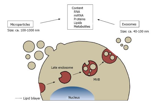
Figure 1 Classification of extracellular vesicles.
Types of extracellular vesicles are distinguished by their mode of generation. Microparticles directly bud off the plasma membrane and measure approximately 100-1000 nm. Exosomes are released by fusion of multivesicular bodies with the plasma membrane and their sizes range between 40-100 nm. Both types of extracellular vesicles can contain RNA, microRNA, proteins, lipids and metabolites. MVBs: Multivesicular bodies.
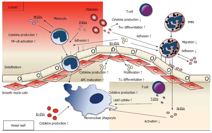
Figure 2 Roles of extracellular vesicles in leukocyte function in the atherosclerotic plaque.
Data on interaction of EVs with neutrophilic granulocytes, monocytes and mononuclear phagocytes and T lymphocytes is summarized. Regarding neutrophilic granuloctes (PMN), platelet EVs (P-EV) promote neutrophil (PMN) adhesion to the endothelium, neutrophil EVs (N-EV) mostly decrease adhesion and migration through the endothelium. Regarding monocytic cells, endothelial (En-EV) and P-EVs promote adhesion to the endothelium. Inside the plaque, En-EVs promote antigen presenting cell (APC) maturation and cytokine production and erythrocyte EVs (Er-EVs), monocyte EVs (M-EVs) and T cell EVs (T-EVs) increase cytokine production. T-EVs also increase lipid uptake. N-EVs suppress activation. Regarding T cells, P-EVs decrease cytokine production, En-EVs promote T cell proliferation and TH1 differentiation. EVs: Extracellular vesicles.
- Citation: Helmke A, von Vietinghoff S. Extracellular vesicles as mediators of vascular inflammation in kidney disease. World J Nephrol 2016; 5(2): 125-138
- URL: https://www.wjgnet.com/2220-6124/full/v5/i2/125.htm
- DOI: https://dx.doi.org/10.5527/wjn.v5.i2.125


